Our Publications
Peer-reviewed papers, books and book chapters advancing computational neurosurgery and brain imaging science.
Our most impactful work
These publications represent the lab's core scientific contributions, from AI-driven tumour segmentation and spectroscopic fingerprinting to fluorescence-guided surgery and the Declaration of Sydney.
Selected recent papers
Browse, filter, and read peer-reviewed articles, conference proceedings, and book chapters and more.
Books and book chapters
Comprehensive monographs and invited chapters authored by lab members, covering computational approaches to neurosurgical disease.

Computational Neurosurgery
Di Ieva, A., Suero Molina, E., Liu, S., & Russo, C. (Eds.) (2024). Computational neurosurgery. (Advances in Experimental Medicine and Biology; Vol. 1462). Springer. https://doi.org/10.1007/978-3-031-64892-2
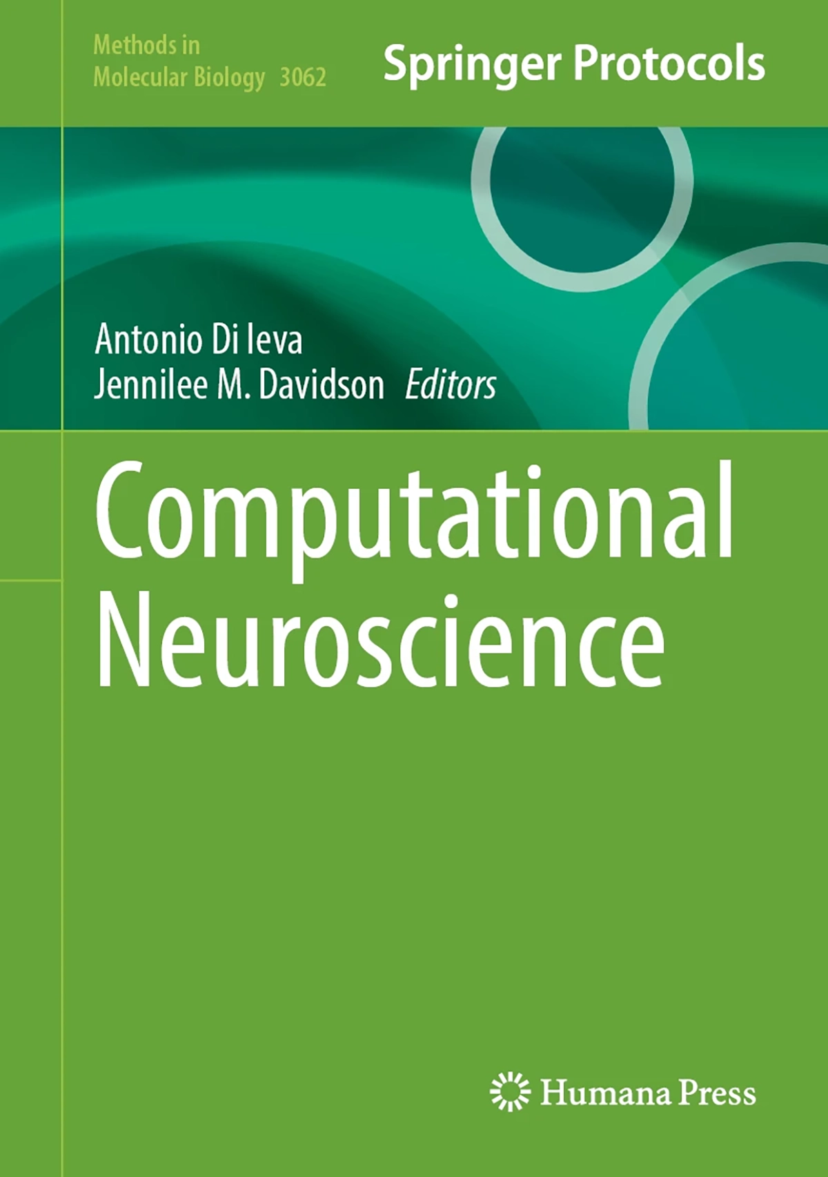
Computational Neuroscience
Di Ieva, A., Davidson, J. (Eds)(2026). Computational neuroscience. Springer. https://link.springer.com/book/9781071654392

The Fractal Geometry of the Brain
Di Ieva, A., (2024). The Fractal Geometry of the Brain. Springer. https://link.springer.com/book/10.1007/978-3-031-47606-8
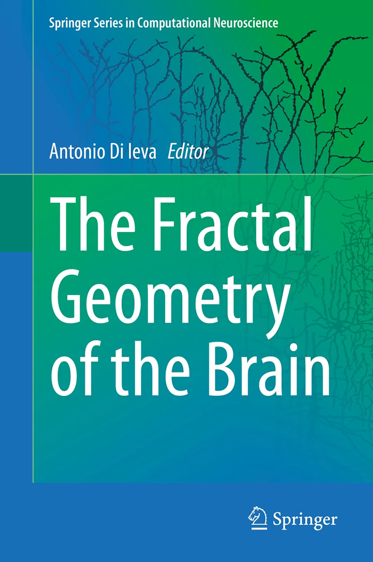
The Fractal Geometry of the Brain
Di Ieva A. (2016). The Fractal Geometry of the Brain. Springer. https://link.springer.com/book/10.1007/978-1-4939-3995-4?page=2
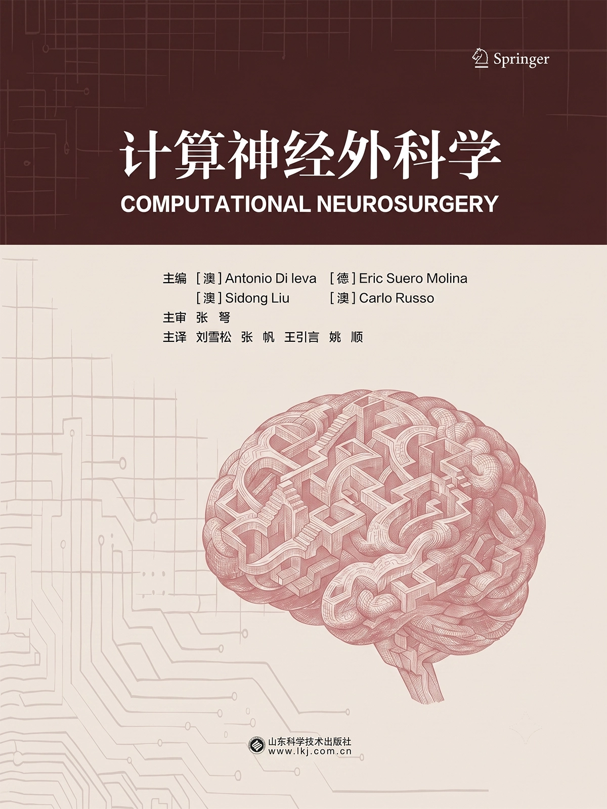
Computational Neurosurgery - Chinese Translation
Di Ieva, A., Suero Molina, E., Liu, S., & Russo, C. (Eds.) (2026). Computational neurosurgery. Springer.
Computational Pipelines for Protein Ubiquitylation Analysis and Prediction
Potapenko, A. et al. (2026). Computational Pipelines for Protein Ubiquitylation Analysis and Prediction. In: Zhang, X., Chen, R., Li, J. (eds) Protein Post-Translational Modifications. Methods in Molecular Biology, vol 3018. Humana, New York, NY. https://doi.org/10.1007/978-1-0716-5166-7_4
The fractal geometry of the brain: an overview
Di Ieva A.. The fractal geometry of the brain: an overview. The fractal geometry of the brain. 2024;:3--13.
Neurosurgery, explainable AI, and legal liability
Matulionyte R., Suero Molina E., Di Ieva A.. Neurosurgery, explainable AI, and legal liability. Computational neurosurgery. 2024;:543--553.
Meta-transfer learning for brain tumor segmentation: within and beyond glioma
Yan S., Liu S., Di Ieva A., Pagnucco M., Song Y.. Meta-transfer learning for brain tumor segmentation: within and beyond glioma. Computational neurosurgery. 2024;:221--230.
Machine and deep learning in hyperspectral fluorescence-guided brain tumor surgery
Suero Molina E., Black D., Xie A., Gill J., Di Ieva A., Stummer W.. Machine and deep learning in hyperspectral fluorescence-guided brain tumor surgery. Computational neurosurgery. 2024;:245--264.
Large language models in neurosurgery
Di Ieva A., Stewart C., Suero Molina E.. Large language models in neurosurgery. Computational neurosurgery. 2024;:177--198.
Fractals, pattern recognition, memetics, and AI: a personal journal in the computational neurosurgery
Di Ieva A.. Fractals, pattern recognition, memetics, and AI: a personal journal in the computational neurosurgery. The fractal geometry of the brain. 2024;:273--283.
Fractals in the neurosciences: a translational geographical approach
Andronache I., Peptenatu D., Ahammer H., Radulovic M., Djuričić G.J., Jelinek H.F., Russo C., Di Ieva A.. Fractals in the neurosciences: a translational geographical approach. The fractal geometry of the brain. 2024;:953--981.
Fractals in neuroimaging
Lahmiri S., Boukadoum M., Di Ieva A.. Fractals in neuroimaging. The fractal geometry of the brain. 2024;:429--444.
Fractals in neuroanatomy and basic neurosciences: an overview
Di Ieva A.. Fractals in neuroanatomy and basic neurosciences: an overview. The fractal geometry of the brain. 2024;:141--147.
Fractal-based analysis of arteriovenous malformations (AVMs)
Di Ieva A., Reishofer G.. Fractal-based analysis of arteriovenous malformations (AVMs). The fractal geometry of the brain. 2024;:413--428.
Fractal-based analysis of histological features of brain tumors
Al-Kadi O.S., Di Ieva A.. Fractal-based analysis of histological features of brain tumors. The fractal geometry of the brain. 2024;:501--524.
Fractal time series: background, estimation methods, and performances
Porcaro C., Moaveninejad S., D’Onofrio V., DiIeva A.. Fractal time series: background, estimation methods, and performances. The fractal geometry of the brain. 2024;:95--137.
Fractal geometry meets computational intelligence: future perspectives
Livi L., Sadeghian A., Di Ieva A.. Fractal geometry meets computational intelligence: future perspectives. The fractal geometry of the brain. 2024;:983--997.
Fractal dimension studies of the brain shape in aging and neurodegenerative diseases
Davidson J.M., Zhang L., Yue G.H., Di Ieva A.. Fractal dimension studies of the brain shape in aging and neurodegenerative diseases. The fractal geometry of the brain. 2024;:329--363.
Fractal dimension analysis in neurological disorders: an overview
Díaz Beltrán L., Madan C.R., Finke C., Krohn S., Di Ieva A., Esteban F.J.. Fractal dimension analysis in neurological disorders: an overview. The fractal geometry of the brain. 2024;:313--328.
Fractal analysis in clinical neurosciences: an overview
Di Ieva A.. Fractal analysis in clinical neurosciences: an overview. The fractal geometry of the brain. 2024;:261--271.
Declaration of computational neurosurgery
Di Ieva A., Suero Molina E., Somerville M.A., Beheshti A., Staartjes V.E., Serra C., Theodore N., Elliott J.M., Wesselink E.O., Russo C., Pilitsis J.G., Bennett C.C., Wu S., Hammond F.M., Lozano A.M., Cusimano M.D., Davidson J.M., Castellano J.F., Okonkwo D.O., Arefan D., Lee C., Zanier O., Da Mutten R., Matula C., Rutka J.T., Pease M., Liu S., Stummer W., Matulionyte R., Yang H., Yuwen C., Cheng X., Fan H., Wang X., Ge Z., Cepeda S., Sheehan J.P., Yang J.Y.M., Hamer R.P., Cohen-Gadol A., Hansford J.R., Savage G., Sowman P.F., Stewart C., Kateb B., Sherif C., Perperidis A., Guller A., Hanft S., D’Amico R.S., Sav A., Cong C., Song Y., Nicolosi F., Wiedmann M.K.H., Barone D.G., Noorani I., Magnussen J., Krieg S.M., Meling T.R., De Ridder D., Lawton M.T., Rosenfeld J.V.. Declaration of computational neurosurgery. Computational neurosurgery. 2024;:11--20.
Cross-Modality Synthesis of T1c MRI from Non-contrast Images Using GANs: Implications for Brain Tumor Research
Tabassum M., Rana P., Suero Molina E., Di Ieva A., Liu S.. Cross-Modality Synthesis of T1c MRI from Non-contrast Images Using GANs: Implications for Brain Tumor Research. Artificial Intelligence in Medicine. 2024;:60–69.
Computational neurosurgery: Foundation
Di Ieva A., Suero Molina E., Liu S., Russo C.. Computational neurosurgery: Foundation. Computational neurosurgery. 2024;:1--8.
Computational fractal-based neurosurgery
Di Ieva A., Davidson J.M., Russo C.. Computational fractal-based neurosurgery. Computational neurosurgery. 2024;:97--105.
Computational fractal-based analysis of MR susceptibility-weighted imaging (SWI) in neuro-oncology and neurotraumatology
Di Ieva A.. Computational fractal-based analysis of MR susceptibility-weighted imaging (SWI) in neuro-oncology and neurotraumatology. The fractal geometry of the brain. 2024;:445--468.
Computational and translational fractal-based analysis in the translational neurosciences: an overview
Di Ieva A.. Computational and translational fractal-based analysis in the translational neurosciences: an overview. The fractal geometry of the brain. 2024;:781--793.
Computational fractal-based analysis of brain tumor microvascular networks
Di Ieva A., Al-Kadi O.S.. Computational fractal-based analysis of brain tumor microvascular networks. The fractal geometry of the brain. 2024;:525--544.
Artificial intelligence, radiomics, and computational modeling in skull base surgery
Suero Molina E., Di Ieva A.. Artificial intelligence, radiomics, and computational modeling in skull base surgery. Computational neurosurgery. 2024;:265--283.
Artificial intelligence in brain tumors
Suero Molina E., Azemi G., Russo C., Liu S., Di Ieva A.. Artificial intelligence in brain tumors. Computational neurosurgery. 2024;:201--220.
Artificial intelligence methods
Liu S., Russo C., Suero Molina E., Di Ieva A.. Artificial intelligence methods. Computational neurosurgery. 2024;:21--38.
Analyzing eye paths using fractals
Newport R.A., Liu S., Di Ieva A.. Analyzing eye paths using fractals. The fractal geometry of the brain. 2024;:827--848.
An experimental verification of neurophysics treatment by executing a multifractal analysis of surface electromyography signals on trapezius, abdominals, and adductor muscles in athletes
Rinzivillo C., Kaleagasioglu F., Casciaro F., Scoppa F., Marvulli R., Ware K., Conte E., Di Ieva A.. An experimental verification of neurophysics treatment by executing a multifractal analysis of surface electromyography signals on trapezius, abdominals, and adductor muscles in athletes. Complexity science in human change. 2024;:81--117.
Robotics for approaches to the anterior cranial fossa
Anokwute M.C., Christodoulides A., Campbell R.G., Harvey R.J., Ieva A.D.. Robotics for approaches to the anterior cranial fossa. Robotics in skull-base surgery. 2023;:35--52.
Publications
Data Processing and Machine Learning Methods to Analyze MALDI-TOF MS Data
MALDI-TOF MS is a rapid, sensitive method for generating complex protein profiles from minimal sample volumes. Although its spectra lack direct peptide sequence information, combining MALDI-TOF output with machine learning enables discrimination of diseases based on intensity patterns. Accurate machine learning model development depends on standardized preprocessing to reduce noise, dimensionality, and bias. This protocol demonstrates two key preprocessing approaches-peak picking and spectral binning-and demonstrates their application in training and testing a machine learning model for the diagnosis of glioblastoma.
Entropy and Fractal Dimensions Application in MALDI-TOF MS Data to Train Classification Models
Matrix-Assisted Laser Desorption/Ionization Time-of-Flight (MALDI-TOF) mass spectrometry generates rich biochemical fingerprints ideal for disease screening. However, its spectral output is highly complex, requiring advanced processing before clinical use. Machine learning excels in learning patterns within this high-dimensional data, but it depends on effective feature extraction. This chapter presents preprocessing strategies for MALDI-TOF spectra and demonstrates the use of fractal dimension and entropy analysis as a feature generation method.
Segmentation-Free Multi-Fractal and Radiomics Response Profiling of Live Unstained Confluent Microglia In Vitro Using Phase-Contrast Microscopy Images
This chapter presents a segmentation-free, cell ensemble-level workflow for detecting treatment-induced responses in confluent microglial cultures in vitro, using standard phase-contrast microscopy and Python-based imaging data analysis. Entire fields of view are analyzed after minimal preprocessing, avoiding fixation, staining, and single-cell segmentation. We implement multifractal, lacunarity, and texture analysis (MFTA) to characterize the microglial monolayers. In parallel, we introduce cell radiomics as the adaptation of radiomics principles to label-free microscopy, extracting a high-dimensional panel of intensity- and texture-based features directly from grayscale images of live cells. Together, MFTA and cell radiomics provide complementary, quantitative readouts that are sensitive to subtle changes in ensemble organization. Statistical comparisons employ false discovery rate control to identify robust discriminative features between conditions, with optional validation by conventional morphometry on a subset of segmented cells. The workflow is demonstrated on an experimental dataset by the successful reveal of the effect of a moderate static magnetic field on human C20 microglia cell line.
Computational Pipeline in Neuroradiomics
Neuroradiomics is a field in neuroimaging that extracts large-scale, quantitative features from central and peripheral nervous systems' images using computational techniques. These features capture intensity distributions, textures, shapes, and spatial patterns. By replacing subjective interpretation with algorithm-driven analysis, neuroradiomics improves objectivity, reproducibility, and consistency, while revealing associations that aid diagnosis, prognosis, and monitoring of neurological and neurosurgical diseases. This chapter presents a step-by-step implementation of the neuroradiomics pipeline, with recommended tools and illustrative Python scripts and command-line snippets provided. The workflow begins with dataset preparation, imaging formats, and computational environments, followed by preprocessing procedures to standardize and normalize neuroimaging data. Segmentation methods are then introduced to define regions of interest, after which radiomic features are extracted using dedicated Python libraries. Subsequent sections demonstrate feature selection, machine learning model development, and performance evaluation, all implemented in reproducible Python environments. Throughout, emphasis is placed on transparency, reproducibility, and practical guidance to support both research and clinical applications. An example implementation is provided to demonstrate practical application and illustrate Python-based workflow implementation with code snippets. By the end of this chapter, readers will have a comprehensive and practical framework for implementing neuroradiomics workflows that would enable them to design, adapt, and apply these methods across diverse neurological applications.
Federated Learning Pipeline for Whole-Slide Image Classification in Digital Neuropathology
Federated learning (FL) has emerged as a powerful paradigm for privacy-preserving, collaborative training of machine learning models, particularly valuable in fields like digital pathology, where data sharing is constrained by institutional policies and regulatory concerns. This chapter presents a reproducible computational pipeline for whole-slide image (WSI) classification in digital neuropathology using a federated learning framework. In addition to outlining the end-to-end implementation, including preprocessing, model training, and deployment, we highlight key challenges specific to applying FL in WSI analysis, such as data heterogeneity and communication efficiency. Detailed code snippets, implementation guidance, and deployment recommendations are provided to support real-world adoption. We hope this chapter serves as a valuable resource for neuropathologists, researchers, and machine learning practitioners aiming to bridge technology and medical science in the evolving landscape of digital neuropathology.
Fractal and Machine Learning Analyses of MALDI-TOF Mass Spectrometry Data in Glioblastoma
Data preprocessing is a critical step in the analysis of matrix-assisted laser desorption/ionization-time-of-flight mass spectrometry (MALDI-TOF MS) spectra for machine learning applications, typically involving steps such as spectra trimming, baseline correction, smoothing, transformation, and peak picking or spectral binning. While traditional approaches focus on protein/peptide peaks as features, this study explores a novel method of feature extraction by treating MALDI-TOF spectra as one-dimensional signal array further processed as time-series data. This study investigates the use of computational fractal-based analysis to assess the complexity of MALDI-TOF spectra. Fractal analysis, previously successful in glioblastoma diagnosis using magnetic resonance imaging, was applied here to proteomics data. By treating each MALDI spectrum as a time series and calculating its fractal dimension using various algorithms, machine learning models were trained to differentiate between glioblastoma patients and controls. We demonstrate that fractal dimensions are sufficient to obtain accurate models for glioblastoma diagnosis, despite still underperforming when compared to the traditional feature extraction method. We also show that fractals can be used as support features to increase model performance. This work highlights the potential and limitations of fractal analysis in proteomics, offering a new perspective for disease diagnosis and broadening the available computational tools for data analysis in mass spectrometry.
Glioma Grade Classification from Structural MRI: A Comparative Transfer Learning Study of Deep Learning Feature Extraction and Machine Learning Classifiers
Background: Accurate histological grading of gliomas, distinguishing low-grade (LGG) from high-grade (HGG) lesions, remains a critical determinant of treatment planning and patient prognosis. This study systematically investigates the optimal combination of structural MRI sequences, pretrained convolutional neural network (CNN) architectures, and supervised classifiers for automated glioma grade classification. Methods: Deep features were extracted from four MRI sequences (T1, contrast-enhanced T1 (T1Gd), T2, and FLAIR) from the BraTS 2023 dataset using five pretrained CNNs: VGG16, ResNet50, DenseNet121, EfficientNetB0, and InceptionV3. Features were refined via LASSO selection and classified using Random Forest, Support Vector Machine, XGBoost, Gradient Boosting, k-Nearest Neighbors (KNNs), and a shallow deep neural network. A patient-level 80/20 training–test partition was employed, with five-fold cross-validation used within the training set for feature selection and hyperparameter optimization. A total of 120 modality–extractor–classifier configurations were benchmarked on the held-out test set. Results: InceptionV3-derived features paired with KNN classifiers consistently yielded superior performance. Based on a TOPSIS multi-criteria ranking integrating accuracy, precision, recall, F1-score, and AUC, the best-performing configuration combined T1Gd features with KNN (closeness coefficient = 0.971; accuracy = 0.975, precision = 0.997, recall = 0.948, F1-score = 0.969, AUC = 0.996). The second-ranked configuration, using T1 features with KNN (closeness coefficient = 0.962), achieved significantly higher accuracy, recall, and F1-score, despite its slightly lower composite ranking. Conclusion: Deep feature extraction using InceptionV3 from T1-weighted MRI, coupled with KNN classification, represents a promising and practical approach for slice-level glioma grading and merits further validation in prospective clinical cohorts.
Computational Pipelines for Protein Ubiquitylation Analysis and Prediction
Ubiquitylation is a crucial posttranslational modification that regulates cellular homeostasis and has been linked to a range of diseases. Identification and analysis of ubiquitylated proteins and ubiquitylation sites have historically been successfully performed by mass spectrometry from in vitro and in vivo samples. However, to address some of the challenges and improve the efficiency of ubiquitylation analysis by mass spectrometry, computational tools have been developed and are becoming increasingly popular. In this chapter, we summarize the available computational tools for predicting ubiquitylation in silico and the common approaches used to enrich ubiquitylation in samples for mass spectrometry and computational analysis. We subsequently provide simple steps for molecular biologists to follow for predicting ubiquitylation sites on proteins in silico, and we provide a computational approach to identify average ubiquitin branch sites across samples from ubiquitin-enriched mass spectrometry data.
Cortical and Subcortical Stimulation for Pain: Network Mechanisms and Therapeutic Implications
Background: Chronic pain affects up to one-third of adults worldwide and remains refractory to conventional therapies in a substantial proportion of patients. Increasing evidence suggests that chronic pain arises from maladaptive interactions across distributed peripheral, spinal, and supraspinal networks rather than dysfunction confined to a single anatomic locus.
Objective: To synthesise animal and human evidence relating to supraspinal pain networks and examine how cortical and subcortical neuromodulation may influence these systems in refractory chronic pain.
Methods: This narrative review integrates findings from electrophysiology, neuroimaging, intracranial stimulation, connectomics, and clinical neuromodulation studies. Evidence is organised around interacting systems involved in chronic pain processing: sensory discriminative, salience and affective valuation, thalamocortical oscillatory, descending modulatory, and interoceptive-predictive.
Results: Chronic pain is associated with abnormalities across distributed sensory, salience, oscillatory, and modulatory systems. Distinct neuromodulation targets engage overlapping but nonidentical network mechanisms: motor cortex stimulation has been associated with modulation of descending inhibitory and thalamocortical systems; somatosensory stimulation may influence sensory gain and oscillatory coupling; anterior cingulate and insular stimulation primarily engages the affective salience and interoceptive systems; and deep brain stimulation provides access to thalamic and brainstem modulatory circuits. Variability and waning efficacy across modalities likely reflect heterogeneous and dynamically evolving network pathology.
Conclusions: A network-guided framework conceptualising chronic pain as a disorder of distributed brain systems supports increasingly personalised, connectivity-informed, multisite, and adaptive neuromodulation strategies. Future approaches may benefit from integrating electrophysiologic and connectomic biomarkers to improve patient selection, target engagement, and durability of analgesia.
BT-CAP: a subcomponent-aware and anatomically constrained data augmentation framework for multi-modal brain tumor MRI segmentation
Background:
Data scarcity and class imbalance remain critical challenges in medical image analysis, particularly for brain tumor MRI segmentation, where subcomponents such as enhancing tumor, non-enhancing tumor, cystic component, and peritumoral edema are underrepresented. Existing augmentation strategies, from classical geometric transforms to GAN-based and diffusion model-based synthesis, either lack subcomponent-level control or require extensive generative model training, limiting their practicality in low-data settings.
Materials and methods:
We propose the Brain Tumor Compositional Augmentation Pipeline (BT-CAP), a subcomponent-aware and anatomically constrained augmentation framework for multi-modal MRI. BT-CAP decomposes tumor subcomponents and recomposes them through a sequence of targeted operations, including isotropic scaling, B-spline deformation, Powell-optimized spatial rearrangement, inpainting, interface smoothing, and constrained edema deformation, applied consistently across all MRI modalities and segmentation masks, producing label-ready augmented volumes without additional annotation. We evaluated augmentation diversity (SSIM, label distribution, intensity variation, centroid displacement) and anatomical plausibility on 50 BraTS-PEDs 2025 cases, yielding 250 augmented volumes, and assessed segmentation performance on the full 256-case dataset using 3-fold cross-validation.
Results:
BT-CAP achieved a mean SSIM of 0.956 ± 0.014 with wide subcomponent volume change ranges and realistic intensity heterogeneity, confirming meaningful structural diversity. By architectural design, all augmented segmentation mask voxels were confined within brain boundaries, and zero overlap between deformed edema and the tumor core was observed across all 250 cases. Segmentation experiments showed mean Dice score improvements of 6%–7% for tumor subcomponents and 2%–3% for tumor core and whole tumor compared to training without compositional augmentation, with a computational cost of approximately 2 min per case on CPU.
Conclusion:
BT-CAP establishes a new class of compositional augmentation methods that deliver anatomically structured, label-ready, and scalable data generation without generative model training. The framework is applicable to any multi-class segmentation task where data scarcity and structural fidelity are critical, and is openly available at https://github.com/dramintavallaii/BT-CAP.
Enhancing Pediatric Brain Tumor Segmentation with Attention-Guided 3D U-Net and a Multi-step Tumor-Aware Compositional Augmentation Pipeline in BraTS 2025
Accurate segmentation of pediatric brain tumors, especially midline and brainstem gliomas, is crucial for neurosurgery and radiotherapy planning, but is hindered by sparse enhancing tumor (ET), infiltrative non-enhancing tumor core (NET), cystic components (CC), and extensive peritumoral edema (ED). We propose an attention-guided 3D U-Net with Multi-Modal and Channel-Wise Attention to enhance feature extraction from multi-parametric MRI (T1, T1-Gd, T2, T2-FLAIR), paired with a novel multi-step tumor-aware compositional augmentation pipeline to simulate tumor variability. Evaluated on the BraTS-PEDs 2025 training set (256 cases) using 3-fold cross-validation, our model achieves robust lesion-wise mean Dice scores: 0.81 ± 0.04 (ET), 0.87 ± 0.05 (NET), 0.77 ± 0.05 (CC), 0.95 ± 0.02 (ED), 0.90 ± 0.03 (WT), and 0.91 ± 0.03 (TC). Hausdorff Distance (95th percentile) values are 57.8 ± 0.6 mm (ET), 13.5 ± 0.7 mm (NET), 63.7 ± 0.8 mm (CC), 6.3 ± 0.4 mm (ED), 8.2 ± 0.5 mm (WT), and 7.1 ± 0.5 mm (TC). Compared to nnU-Net, our model improves WT and TC Dice by 2% and 6%, respectively, driven by attention mechanisms and augmentation. Ablation studies show a 6–7% Dice drop without augmentation, highlighting its role in generalizability. Increased WT and TC Dice performance directly supports more accurate radiotherapy margin definition and prognostic modeling for survival prediction, highlighting the clinical utility of our approach. Despite remaining challenges with sparse ET and irregular cystic components, our framework demonstrates robustness and scalability, paving the way for translation into clinical neuro-oncology workflows.
Distinctive features of the tumor and immune microenvironment in glioblastoma
Immune checkpoint inhibitors (ICI) have improved outcomes for asymptomatic patients with melanoma brain metastases (MBM) but have delivered limited benefit in glioblastoma (GBM). Defining context-dependent features of ICI response is critical for tailoring effective therapeutic strategies and improving GBM patient outcomes. Multiparameter flow cytometry analyses of immune composition and activation status were performed on 110 tumor cell suspensions obtained from cavitron ultrasonic surgical aspirates (CUSA) specimens (n = 40), and surgically resected GBM (n = 21) and MBM (n = 49) tumors. GBM tumors exhibited marked phenotypic heterogeneity, frequently co-expressing stem-like and differentiation markers. GBM stemness correlated with increased macrophage infiltration and reduced T cell abundance, implicating plasticity in the establishment of an immunosuppressive microenvironment. Compared to MBM, GBM showed a marked paucity of T cells, an enrichment of myeloid cells, downregulated MHC-I, and elevated PD-L2 expression on tumor cells. Multiplex immunostaining performed on a subset of GBM samples, confirmed myeloid predominance and indicated pronounced spatial heterogeneity. However, the presence of antigen-experienced T cells and preserved IFNγ signaling indicates a latent potential for immune engagement. These findings provide a rationale for ICI-based combinations designed to restore effective anti-tumor immunity in GBM.
Quantifying the development of visual expertise in medical image interpretation using fractal eye-gaze metrics
This study presents a fractal-based framework for quantifying visual expertise using eye-gaze data. A longitudinal experiment was conducted across three consecutive semesters of the medical program at Macquarie University, where the eye movements of 13 medical students were recorded with a high-resolution eye tracker while they viewed pathological images from multiple imaging modalities. In addition, eye-tracking data from 13 consultant neurosurgeons were included as expert reference data to assess whether longitudinal changes in students’ approach mirror expert-like visual behavior. Three metrics were computed: the two-and three-dimensional fractal dimensions of eye-gaze to capture spatiotemporal complexity of the eye-gaze data, and a fractal-dimension–based correlation to quantify the relationship between gaze patterns and stimulus structure. These metrics were combined into a composite Fractal Eye-Gaze Expertise Index (FEI). The results show systematic reductions in fractal complexity across sessions, indicating increasingly structured and efficient gaze strategies, with trends approaching those of experts. Crucially, conventional fixation-duration–based measures failed to consistently capture this progression, unlike the proposed fractal metrics. Statistical analyses confirm significant session-related effects for all metrics, and receiver operating characteristic curve (ROC) analysis shows that the FEI effectively discriminates longitudinal differences in visual expertise. This work demonstrates that fractal-based eye-gaze modelling provides an objective and scalable approach to characterizing visual expertise.
Computational neurosurgery - Chinese Translation
This comprehensive and authoritative reference presents the state-of-the-art computational methods applied to the field of neurosurgery. The book brings together leading neuroscientists, neurosurgeons, mathematicians, computer scientists, engineers, ethicists and lawyers, to open the new frontier of computational neurosurgery to a broad audience interested in the translational field of the application of computational models, such as deep learning, to the study of the brain and the practical applications of neurosurgery. The focus is primarily clinical, and there is a solid foundation of research aspects. With forewords by Michael L.J. Apuzzo and Enrico Coiera, the book is organized into 2 sections: (1) tenets of computational modeling, artificial intelligence, computational analysis, and analysis software; (2) computational neurosurgery applications, including neurodiagnostics, neuro-oncology, vascular neurosurgery, all the neurosurgical disciplines, surgical approaches, intraoperative applications, and ethics and legal aspects
The Declaration of Sydney: Ethical, Legal, and Professional Foundations of Computational Neurosurgery
Artificial intelligence (AI), data-driven methods, and emerging neurotechnologies are increasingly shaping how neurosurgery is practised, studied, and taught. Within this evolving landscape, computational neurosurgery has emerged as a transdisciplinary field that integrates computation, artificial and augmented intelligence, data science, and neurotechnology across the neurosurgical care pathway, encompassing applications in diagnosis, risk stratification and screening, clinical decision support, surgical planning, intraoperative support, postoperative management, research, education, and training, with the overarching aim of improving fundamental understanding, clinical decision-making, and patient outcomes. Computational methodologies have been shown to, or at least promise to, influence clinical judgment throughout neurosurgical care. While such developments hold considerable potential for enhancing precision and expanding access to care, they also introduce ethical, legal, and societal questions that are not readily addressed by generic approaches to AI governance. Neurosurgery occupies a particularly sensitive position in this context, given its direct engagement with the brain as the substrate of consciousness, identity, and agency.
In response to these challenges, an international and multidisciplinary task force was convened in association with the first World Conference of Computational Neurosurgery (WCCNS, Sydney, Australia, 13-15 February 2026). Its goal was to develop a shared ethical framework and embed it in neurosurgical practice, to guide the responsible use of AI and computational methods in neurosurgery and other complementary disciplines. Working through an iterative process of deliberation, collaborative drafting, and explicit documentation of consensus and disagreement, the task force produced two complementary outcomes: a concise public pledge, the Declaration of Sydney, and the present white paper, which elaborates the ethical and conceptual foundations underlying that pledge.
This white paper examines the emergence of computational neurosurgery as both a clinical and epistemic shift, and explores its implications for governance, data stewardship, clinical integration, education, equity, sustainability, and long-term oversight. With a patient-centred approach, the Declaration of Sydney affirms the primacy of patients, respect for human dignity, professional accountability, and transparency, while acknowledging the need for governance frameworks capable of adapting to evolving technologies and societal expectations. Taken together, the goal of the Declaration and the analysis presented in this Whitepaper is to benefit patients needing neurosurgical care, support responsible innovation, encourage transdisciplinary engagement, and contribute to the ethical maturation of computational neurosurgery as a global field. In such a perspective, the Declaration of Sydney emerges as a global framework for responsible computational neurosurgery, defining the articles and ethical principles neurosurgeons should abide by when using AI and computational techniques.
Computational Neuroscience
This detailed volume explores practical and reproducible computational techniques and pipelines that span the translational neurosciences continuum. The first section of the book focuses on experimental platforms that generate high-dimensional biological data and the computational methodologies required to transform raw signals into mechanistic insights, while the second half transitions from bench to bedside, illustrating how computational methodologies reshape diagnosis, prognosis, and mechanistic inference across the whole pathophysiological spectrum of the nervous system, from normal brain function to neurological diseases. Written for the highly successful Methods in Molecular Biology series, each chapter provides step-by-step workflows, code snippets, tool recommendations, and the practical guidance needed for positive results.
What Can We Count On? Performance of Microplate Cell Counting Assays in 2D Monolayer and 3D ECM-based In Vitro Tumour Models
Collaborative learning across medical institutions is essential for building robust and generalisable digital pathology models. Federated learning (FL) enables collaboration without centralising data, yet its adoption is limited by high communication costs, model heterogeneity, and privacy concerns. We propose Federated Deep Feature Prompting (FedDFP), an efficient FL framework tailored for heterogeneous clinical environments. FedDFP introduces lightweight, client-specific learnable prompts applied to patch-level embeddings from whole-slide images. By sharing only these compact prompts, FedDFP reduces communication overhead by over 99.9% compared to standard FL while improving classification accuracy. Extensive experiments on TCGA-IDH, CAMELYON16 and CAMELYON17 show that FedDFP consistently outperforms standard and personalised FL baselines, achieving mean AUC gains of 0.11-0.13 over local-only training and up to 0.10 over the strongest federated methods. FedDFP also converges 2-4× faster and remains effective across diverse feature extractors and multiple-instance learning architectures, demonstrating scalability, flexibility and privacy-aware collaboration.
The trust gap in generative medical imaging: evidence, risks, and a roadmap toward responsible adoption
Generative artificial intelligence produces radiological images and clinical text with a level of realism that suggests broad utility in medical imaging. Early studies show promise in targeted data augmentation, assistive image enhancement and reconstruction, cautious cross-modality synthesis, and report drafting. Yet realism is not reliability. Generative systems may introduce or omit findings, breach basic physics or anatomy, fail under data distribution shifts, leak private information, or degrade when synthetic data re-enters training pipelines. Risks from human factors such as automation bias, provenance blindness, and susceptibility to manipulation further widen the gap between plausible output and clinical truth. In this review, we focus on narrowing that gap. To support safe translation, we extend prior evaluation schemes into a three-tier evaluation framework, progressing from pixel- and physics-level checks to anatomy-level realism and task-level clinical utility. Building on this, we outline a five-layer “trustworthiness stack” spanning data governance, physics-informed model design, multi-tier evaluation with continuous monitoring, human-centered interfaces, and institutional oversight. Finally, we provide stakeholder-specific recommendations for implementing generative imaging systems as constrained, auditable, continuously monitored components of clinical practice that augment—but do not replace—expert judgment.
Slide-aware deep feature prompting for enhanced Whole Slide Image classification
The advent of Whole Slide Imaging (WSI) has revolutionised digital pathology by enabling computational analysis of gigapixel-scale images. To handle their large size, most deep learning models divide WSIs into patches and apply Multiple Instance Learning (MIL) for slide-level classification. However, MIL models often depend on pre-trained feature extractors, resulting in domain gaps between natural and pathological images. Parameter-Efficient Fine-Tuning (PEFT) via visual prompting has emerged to bridge this gap with minimal overhead. Nevertheless, existing visual prompts are typically attached at the image level and tightly coupled with specific architectures such as CNNs or ViTs, limiting generalisability and scalability in WSI tasks. To overcome these limitations, we propose Slide-aware Deep Feature Prompt (S-DFP), a novel visual prompting method which derives task-specific information directly from feature embeddings and is initialised with slide-specific cues, thereby enhancing compatibility with diverse feature extractors and MIL frameworks. Experiments on four benchmark datasets, CAMELYON16, BRIGHT, TCGA-IDH, and UniToPath, demonstrate that S-DFP consistently boosts MIL model performance by 2–5% in AUC while introducing less than 0.02% additional parameters. Furthermore, when integrated with recent pathology foundation models, S-DFP yields additional performance gains. The code is publicly available at S-DFP.
Prediction of remission in cushing disease using artificial intelligence: a systematic review and meta-analysis
Predicting remission in Cushing’s disease (CD) following transsphenoidal surgery (TSS) is crucial for improving treatment strategies and patient outcomes. This systematic review and meta-analysis aimed to evaluate the application of Artificial Intelligence (AI) algorithms in forecasting remission outcomes, integrating diverse clinical, biochemical, and imaging data to enhance predictive accuracy. A comprehensive search was conducted across PubMed, Web of Science, Embase, Scopus, and Google Scholar databases up to December 2024, adhering to PRISMA guidelines. Out of 1,571 records identified, five studies involving 1,938 patients met the inclusion criteria. The pooled sensitivity and specificity of Machine learning (ML) models were 0.93 [95% CI: 0.65–0.99] and 0.78 [95% CI: 0.50–0.93], respectively, with the diagnostic score was 3.8 [95% CI: 0.86–6.74] and diagnostic odds ratio (DOR) of 44.79 [95% CI: 2.37–846.66]. The positive diagnostic likelihood ratio (DLR) was 4.23 [95% CI: 1.39–12.86], while the negative DLR was 0.09 [95% CI: 0.01–0.67]. The area under the curve (AUC) was 0.91. These results underscore the significant potential of AI algorithms in enhancing clinical decision-making and improving the prediction of remission in CD. However, methodological heterogeneity and the lack of external validation across studies call for standardized approaches to ensure broader applicability and reliability of these models in clinical settings.
Explainable brain tumor detection with eye fixation density map alignment
Deep learning-based object detectors have demonstrated strong performance in localizing brain tumors on magnetic resonance imaging (MRI). However, clinical adoption requires not only accurate predictions but also trustworthy explanations that align with expert reasoning. Saliency-based explainable AI (XAI) methods visualize where a model focuses its attention. Despite this, their alignment with expert visual attention and robustness to benign image variations remains insufficiently studied in neuroimaging. To address this gap, we propose a gaze-informed evaluation framework that quantitatively assesses the interpretability of a highperforming explainable AI model using expert eye-tracking data. Our Method compares saliency maps from two state-of-the-art object detectors against expert eye-tracking fixation density maps (ETFDs) collected from experienced neurosurgeons. We employ two complementary metrics: Normalized Cross-Correlation (NCC) for global saliency similarity and Normalized Scanpath Saliency (NSS) for point-wise fixation correspondence. The proposed approach offers valuable insights into the clinical trustworthiness of saliency-based explanations in neuroimaging.
Characterizing visual neurosurgical expertise in brain MRI visualization using eye-tracking and 3D fractal dimension analysis
Eye-tracking has been utilized to characterize visual behavior in medical image visualization and interpretation, yet neurosurgeons remain underrepresented. Characterizing neurosurgery-specific visual expertise is important for understanding expert search strategies, informing training, and developing computational models. This study examined gaze behavior in naïve observers (Np = 29), neurosurgery registrars (Np = 16), and consultant neurosurgeons (Np = 24), viewing normal (Np = 20) and pathological (Np = 19) brain MR images under a free-viewing paradigm. To capture expertise-related characteristics, we analyzed two features at each fixation location: (i) fixation duration, reflecting temporal allocation of visual attention, and (ii) three-dimensional fractal dimension (3DFD) around each fixation location, quantifying local structural complexity. To assess pathological-type effects, we grouped similar pathologies into five stimulus groups. Linear mixed-effects modelling revealed systematic expertise-related differences, with experts exhibiting longer fixation durations in pathological stimulus groups and pathology-type-dependent complexity sampling. Combined fixation duration and 3DFD features captured complementary aspects of visual expertise, improving Random Forest classifier’s accuracy (>93%) compared to individual features, for all five stimulus groups. These findings highlight neurosurgery-specific markers of visual expertise and demonstrate that combining behavioral and image-derived features could underpin computational models and training tools that emulate expert-level strategies in neurosurgical image interpretation. Future work should evaluate its applicability to other medical domains.
Towards a Multimodal MRI-Based Foundation Model for Multi-Level Feature Exploration in Segmentation, Molecular Subtyping, and Grading of Glioma
Accurate, noninvasive glioma characterization is crucial for effective clinical management. Traditional methods, dependent on invasive tissue sampling, often fail to capture the spatial heterogeneity of the tumor. While deep learning has improved segmentation and molecular profiling, few approaches simultaneously integrate tumor morphology and molecular features. Foundation deep learning models, which learn robust, task-agnostic representations from large-scale datasets, hold great promise but remain underutilized in glioma imaging biomarkers. We propose the Multi-Task SWIN-UNETR (MTS-UNET) model, a novel foundation-based framework built on the BrainSegFounder model, pretrained on large-scale neuroimaging data. MTS-UNET simultaneously performs glioma segmentation, histological grading, and molecular subtyping (IDH mutation and 1p/19q co-deletion). It incorporates two key modules: Tumor-Aware Feature Encoding (TAFE) for multi-scale, tumor-focused feature extraction and Cross-Modality Differential (CMD) for highlighting subtle T2-FLAIR mismatch signals associated with IDH mutation. The model was trained and validated on a diverse, multi-center cohort of 2,249 glioma patients from seven public datasets. MTS-UNET achieved a mean Dice score of 84% for segmentation, along with AUCs of 90.58% for IDH mutation, 69.22% for 1p/19q co-deletion prediction, and 87.54% for grading, significantly outperforming baseline models (p<=0.05). Ablation studies validated the essential contributions of the TAFE and CMD modules and demonstrated the robustness of the framework. The foundation-based MTS-UNET model effectively integrates tumor segmentation with multi-level classification, exhibiting strong generalizability across diverse MRI datasets. This framework shows significant potential for advancing noninvasive, personalized glioma management by improving predictive accuracy and interpretability.
Synthetic O-(2-18F-fluoroethyl)-L-tyrosine-positron emission tomography generation and hotspot prediction via preoperative MRI fusion of gliomas lacking radiographic high-grade characteristics
Background: Limited amino acid availability for positron emission tomography (PET) imaging hinders therapeutic decision-making for gliomas without typical high-grade imaging features. To address this gap, we evaluated a generative artificial intelligence (AI) approach for creating synthetic O-(2-18F-fluoroethyl)-l-tyrosine ([18F]FET)-PET and predicting high [18F]FET uptake from magnetic resonance imaging (MRI). Methods: We trained a deep learning (DL)-based model to segment tumors in MRI, extracted radiomic features using the Python PyRadiomics package, and utilized a Random Forest classifier to predict high [18F]FET uptake. To generate [18F]FET-PET images, we employed a generative adversarial network framework and utilized a split-input fusion module for processing different MRI sequences through feature extraction, concatenation, and self-attention. Results: We included magnetic resonance imaging (MRI) and PET images from 215 studies for the hotspot classification and 211 studies for the synthetic PET generation task. The top-performing radiomic features achieved 80% accuracy for hotspot prediction. From the synthetic [18F]FET-PET, 85% were classified as clinically useful by senior physicians. Peak signal-to-noise ratio analysis indicated high signal fidelity with a peak at 40 dB, while structural similarity index values showed structural congruence. Root mean square error analysis demonstrated lower values below 5.6. Most visual information fidelity scores ranged between 0.6 and 0.7. This indicates that synthetic PET images retain the essential information required for clinical assessment and diagnosis. Conclusion: For the first time, we demonstrate that predicting high [18F]FET uptake and generating synthetic PET images from preoperative MRI in lower-grade and high-grade glioma are feasible. Advanced MRI modalities and other generative AI models will be used to improve the algorithm further in future studies.
Source-free few-shot segmentation for rarer brain tumors
Since the inception of BraTS challenge, a series of methods has been developed for brain tumor segmentation over the past years. Although these methods achieved promising results, they mostly focus on glioma segmentation, largely due to their relatively high incidence. These fully-supervised methods may not be applicable as they rely on abundant labeled data, which is intrinsically inaccessible for rarer types of brain tumors. Data-efficient transfer learning approaches like few-shot learning and domain adaptation assume full access to source data, which may not be feasible in real-life scenarios due to privacy and confidentiality concerns. In this work, we propose a new source-free few-shot learning framework for rarer brain tumor segmentation that adapts source model trained on gliomas to other less common brain tumors such as meningioma, metastasis and pediatric tumors with only a few labeled target data. The proposed framework follows a dual-branch prototypes learning structure that harmonize preservation of common knowledge from source class and learning new features from target. We show that our method gains a 6% increase in Dice score over representative source-free domain adaptation methods, and achieves comparable performance against its fully-supervised counterpart.
Radiomic Fingerprinting of the Peritumoral Edema in Brain Tumors
Background: Radiomics is a quantitative approach to medical imaging, aimed to extract features into large datasets. By using artificial intelligence (AI) methodologies, large radiomic data can be analysed and translated into meaningful clinical applications. In rhinology, there is heavy reliance on computed tomography (CT) imaging of the paranasal sinus for diagnostics and assessment of treatment outcomes. Currently, there is an emergence of literature detailing radiomics use in rhinology. Objective: This systematic review aims to assess the current techniques used to analyze radiomic data from paranasal sinus CT imaging. Methods: A systematic search was performed using Ovid MEDLINE and EMBASE databases from January 1, 2019 until March 16, 2024 using the Preferred Reporting Items for Systematic Reviews and Meta-analyses (PRISMA) checklist and Cochrane Library Systematic Reviews for Diagnostic and Prognostic Studies. The QUADAS-2 and PROBAST tools were utilized to assess risk of bias. Results: Our search generated 1456 articles with 10 articles meeting eligibility criteria. Articles were divided into 2 categories, diagnostic (n = 7) and prognostic studies (n = 3). The number of radiomic features extracted ranged 4 to 1409, with analysis including non-AI-based statistical analyses (n = 3) or machine learning algorithms (n = 7). The diagnostic or prognostic utility of radiomics analyses were rated as excellent (n = 3), very good (n = 2), good (n = 2), or not reported (n = 3) based upon area under the curve receiver operating characteristic (AUC-ROC) or accuracy. The average radiomics quality score was 36.95%. Conclusion: Radiomics is an evolving field which can augment our understanding of rhinology diseases, however there are currently only minimal quality studies with limited clinical utility.
Predicting intraoperative 5-ALA-induced tumor fluorescence via MRI and deep learning in gliomas with radiographic lower-grade characteristics
Purpose: Lower-grade gliomas typically exhibit 5-aminolevulinic acid (5-ALA)-induced fluorescence in only 20-30% of cases, a rate that can be increased by doubling the administered dose of 5-ALA. Fluorescence can depict anaplastic foci, which can be precisely sampled to avoid undergrading. We aimed to analyze whether a deep learning model could predict intraoperative fluorescence based on preoperative magnetic resonance imaging (MRI).Methods: We evaluated a cohort of 163 glioma patients categorized intraoperatively as fluorescent (n = 83) or non-fluorescent (n = 80). The preoperative MR images of gliomas lacking high-grade characteristics (e.g., necrosis or irregular ring contrast-enhancement) consisted of T1, T1-post gadolinium, and FLAIR sequences. The preprocessed MRIs were fed into an encoder-decoder convolutional neural network (U-Net), pre-trained for tumor segmentation using those three MRI sequences. We used the outputs of the bottleneck layer of the U-Net in the Variational Autoencoder (VAE) as features for classification. We identified and utilized the most effective features in a Random Forest classifier using the principal component analysis (PCA) and the partial least square discriminant analysis (PLS-DA) algorithms. We evaluated the performance of the classifier using a tenfold cross-validation procedure.Results: Our proposed approach's performance was assessed using mean balanced accuracy, mean sensitivity, and mean specificity. The optimal results were obtained by employing top-performing features selected by PCA, resulting in a mean balanced accuracy of 80% and mean sensitivity and specificity of 84% and 76%, respectively.Conclusions: Our findings highlight the potential of a U-Net model, coupled with a Random Forest classifier, for pre-operative prediction of intraoperative fluorescence. We achieved high accuracy using the features extracted by the U-Net model pre-trained for brain tumor segmentation. While the model can still be improved, it has the potential for evaluating when to administer 5-ALA to gliomas lacking typical high-grade radiographic features.
Multi-sequence MRI to multi-tracer PET generation via diffusion model
Magnetic Resonance Imaging (MRI) and Positron Emission Tomography (PET) are essential tools for diagnosing brain diseases. However, PET image acquisition is typically associated with high costs and significant radiation exposure to patients. Recent works have leveraged generative models to synthesise PET images from MRI scans. However, previous methods undergo training multiple times to accommodate the varying tracers of PET images, leading to limited flexibility. Additionally, they rely on simple concatenation when combining multiple MRI sequences, overlooking the correlations between these sequences. To address these limitations, this paper proposes a novel Diffusion Model that accepts multiple MRI sequences as inputs and generates PET images with various tracers. The model integrates an image encoder that processes multiple MRI sequences with cross attention and a categorical embedding that encodes the specific tracer information to guide the PET image generation. Experimental results on the public Alzheimer's Disease Neuroimaging Initiative (ADNI) dataset demonstrate that our method offers higher flexibility and superior performance in PET image generation compared to the current state-of-the-art. The source code is available at: https://github.com/ZJohnWenjin/MTGD.git
FoundBioNet: A Foundation-Based Model for IDH Genotyping of Glioma from Multi-parametric MRI
Brain disease diagnosis and treatment planning rely on complementary information from multiple MRI modalities. Compared to routine modalities (RM) such as T1, T2, and FLAIR, modalities like DWI and T1ce provide unique diagnostic information but are less commonly used due to longer scan times, higher costs, or the need for contrast agents. To mitigate this, multi-modal MRI synthesis methods are proposed to generate advanced MRIs from routine MRIs. However, in clinical practice, missing modality is a known issue in MRI generation which degrades the synthesis quality. Existing methods typically use shared encoders and masking strategies to compensate for missing modality. However, as the number of missing modalities increases, it becomes harder to capture the inter-modal correlations, causing a sharp performance drop. To address this, we propose the Feature Mapping and Merging Diffusion Model (FMM-Diff). Instead of using a shared encoder, we introduce dedicated mapping encoders for each modality. When a modality is missing, its latent representation is inferred from the available ones via its dedicated encoder. This ensures complete latent representations, allowing the Merge Module to selectively extract and fuse inter-modal correlations, significantly improving synthesis performance. Evaluated on two public MRI datasets, including CGGA and BraTS2021, FMM-Diff not only outperforms the state-of-the-art models by 4.35% in terms of Structural Similarity Index Measure (SSIM) while demonstrating exceptional stability, with less than a 1.0% SSIM drop, which is significantly lower than the 2.0–3.45% drop observed with other methods, across various missing modality scenarios. The source code will be available at: https://github.com/ZJohnWenjin/FMMDIFF.git.
Eye-Guided Multimodal Fusion: Toward an Adaptive Learning Framework Using Explainable Artificial Intelligence
In recent years, numerous algorithms have emerged for the segmentation of brain tumors, driven by advancements in deep learning techniques, where the objective is to identify and delineate various tumor sub-regions. While deep learning models like nnUNet have shown promising results in glioma segmentation, their effectiveness in segmenting other brain tumor subtypes, such as meningiomas and metastases, remains uncertain, especially when the available dataset lacks representative examples. To address this challenge, we propose a meta-transfer learning approach, which involves fine-tuning the nnUNet model on datasets containing meningiomas and metastases while leveraging the knowledge acquired from glioma segmentation. This approach aims to enhance the adaptability of nnUNet, allowing it to generalize better to diverse brain tumor types and potentially improving the accuracy of diagnosis and treatment planning for patients with meningiomas and metastases. Our proposed method significantly improves segmentation performance, achieving Dice coefficients of 0.8621 ± 0.2413 for Whole Tumor (WT) in meningiomas and 0.8141 ± 0.0562 for WT in metastases. These results set a new benchmark in brain tumor segmentation and pave the way for more robust and generalizable medical image analysis tools.
Diagnostic Accuracy of Deep Learning Models in Predicting Glioma Molecular Markers: A Systematic Review and Meta-Analysis
Background/Objectives
Integrating deep learning (DL) into radiomics offers a noninvasive approach to predicting molecular markers in gliomas, a crucial step toward personalized medicine. This study aimed to assess the diagnostic accuracy of DL models in predicting various glioma molecular markers using MRI.
Methods
Following PRISMA guidelines, we systematically searched PubMed, Scopus, Ovid, and Web of Science until 27 February 2024 for studies employing DL algorithms to predict gliomas’ molecular markers from MRI sequences. The publications were assessed for the risk of bias, applicability concerns, and quality using the QUADAS-2 tool and the radiomics quality score (RQS). A bivariate random-effects model estimated pooled sensitivity and specificity, accounting for inter-study heterogeneity.
Results
Of 728 articles, 43 were qualified for qualitative analysis, and 30 were included in the meta-analysis. In the validation cohorts, MGMT methylation had a pooled sensitivity of 0.74 (95% CI: 0.66–0.80) and a pooled specificity of 0.75 (95% CI: 0.65–0.82), both with significant heterogeneity (p = 0.00, I2 = 80.90–84.50%). ATRX and TERT mutations had a pooled sensitivity of 0.79 (95% CI: 0.67–0.87) and 0.81 (95% CI: 0.72–0.87) and a pooled specificity of 0.85 (95% CI: 0.78–0.91) and 0.70 (95% CI: 0.61–0.77), respectively. Meta-regression analyses revealed that significant heterogeneity was influenced by data sources, MRI sequences, feature extraction methods, and validation techniques.
Conclusions
While the DL models show promising prediction accuracy for glioma molecular markers, variability in the study settings complicates clinical translation. To bridge this gap, future efforts should focus on harmonizing multi-center MRI datasets, incorporating external validation, and promoting open-source studies and data sharing.
Development of an open-source algorithm for automated segmentation in clinician-led paranasal sinus radiologic research
Objective: Artificial Intelligence (AI) research needs to be clinician led; however, expertise typically lies outside their skill set. Collaborations exist but are often commercially driven. Free and open-source computational algorithms and software expertise are required for meaningful clinically driven AI medical research. Deep learning algorithms automate segmenting regions of interest for analysis and clinical translation. Numerous studies have automatically segmented paranasal sinus computed tomography (CT) scans; however, openly accessible algorithms capturing the sinonasal cavity remain scarce. The purpose of this study was to validate and provide an open-source segmentation algorithm for paranasal sinus CTs for the otolaryngology research community. Methods: A cross-sectional comparative study was conducted with a deep learning algorithm, UNet++, modified for automatic segmentation of paranasal sinuses CTs and “ground-truth” manual segmentations. A dataset of 100 paranasal sinuses scans was manually segmented, with an 80/20 training/testing split. The algorithm is available at https://github.com/rheadkaul/SinusSegment. Primary outcomes included the Dice similarity coefficient (DSC) score, Intersection over Union (IoU), Hausdorff distance (HD), sensitivity, specificity, and visual similarity grading. Results: Twenty scans representing 7300 slices were assessed. The mean DSC was 0.87 and IoU 0.80, with HD 33.61 mm. The mean sensitivity was 83.98% and specificity 99.81%. The median visual similarity grading score was 3 (good). There were no statistically significant differences in outcomes with normal or diseased paranasal sinus CTs. Conclusion: Automatic segmentation of CT paranasal sinuses yields good results when compared with manual segmentation. This study provides an open-source segmentation algorithm as a foundation and gateway for more complex AI-based analysis of large datasets. Level of Evidence: 3.
Artificial intelligence for brain neuroanatomical segmentation in magnetic resonance imaging: a literature review
Purpose: This literature review aims to synthesise current research on the application of artificial intelligence (AI) for the segmentation of brain neuroanatomical structures in magnetic resonance imaging (MRI). Methods: A literature search was conducted using the databases Embase, Medline, Scopus, and Web of Science, and captured articles were assessed for inclusion in the review. Data extraction was performed for the summary of the AI model used, and key findings of each article, advantages and disadvantages were identified. Results: Following full-text screening, 21 articles were included in the review. The review covers models for segmentation models applied to the whole brain, cerebral cortex, subcortical structures, the cerebellum, blood vessels, perivascular spaces, and the ventricles. Accuracy of segmentation was generally high, particularly for segmenting neuroanatomical structures in healthy cohorts. Conclusion: The use of AI for automatic brain segmentation is generally highly accurate and fast for all regions of the human brain. Challenges include robustness to anatomical variability and pathology, largely due to difficulties with accessing sufficient training data.
Adaptive Clustering for EGFR Amplification Prediction in Glioblastoma: A Variational Autoencoder-Dirichlet Bayesian Gaussian Approach
Introduction: Pain management in patients with complete spinal cord injury is complex. Case Report: We report a successful case of managing neuropathic, phantom limb, and back pain below the level of spinal cord injury (T5 American Spinal Injury Association [ASIA] A) using a 10 kHz high-frequency spinal cord stimulator (SCS) over a 6-month follow-up period. Conclusion: The effectiveness of this approach may be attributed to its ability to modulate supraspinal pain processing, allowing for targeted relief of various pain mechanisms below the level of injury.
When Eye-Tracking Meets Machine Learning: A Systematic Review on Applications in Medical Image Analysis
Eye-gaze tracking research offers significant promise in enhancing various healthcare-related tasks, above all in medical image analysis and interpretation. Eye tracking, a technology that monitors and records the movement of the eyes, provides valuable insights into human visual attention patterns. This technology can transform how healthcare professionals and medical specialists engage with and analyze diagnostic images, offering a more insightful and efficient approach to medical diagnostics. Hence, extracting meaningful features and insights from medical images by leveraging eye-gaze data improves our understanding of how radiologists and other medical experts monitor, interpret, and understand images for diagnostic purposes. Eye-tracking data, with intricate human visual attention patterns embedded, provides a bridge to integrating artificial intelligence (AI) development and human cognition. This integration allows novel methods to incorporate domain knowledge into machine learning (ML) and deep learning (DL) approaches to enhance their alignment with human-like perception and decision-making. Moreover, extensive collections of eye-tracking data have also enabled novel ML/DL methods to analyze human visual patterns, paving the way to a better understanding of human vision, attention, and cognition. This systematic review investigates eye-gaze tracking applications and methodologies for enhancing ML/DL algorithms for medical image analysis in depth.
The Fractal Geometry of the Brain, 2nd Edition
The new edition of the highly popular, The Fractal Geometry of the Brain, reviews the most intriguing applications of fractal analysis in neuroscience with a focus on current and future potential, limits, advantages, and disadvantages. It brings an understanding of fractals to clinicians and researchers even if they do not have a mathematical background, and it serves as a valuable tool for teaching the translational applications of computational fractal-based models to both students and scholars. As a consequence of the novel research developed at Professor Di Ieva's laboratory and other centers around the world, the second edition will explore the use of computational fractal-based analysis in many clinical disciplines and different fields of research, including neurology and neurosurgery, neuroanatomy and psychology, magnetoencephalography (MEG), eye-tracking devices (for the fractal computational characterization of "scanpaths"),deep learning in image analysis, radiomics for the characterization of brain MRIs, characterization of neuropsychological and psychiatric diseases and traits, signal complexity analysis in time series, and functional MRI, amongst others.
Use of artificial intelligence in the prediction of Chiari malformation type 1 recurrence after posterior fossa decompressive surgery
Purpose The purpose of this study was to train a deep learning-based method for the prediction of postoperative recurrence of symptoms in Chiari malformation type 1 (CM1) patients undergoing surgery. Studies suggest that certain radiological and clinical features do exist in patients with treatment failure, though these are inconsistent and poorly defined. Methodology This study was a retrospective cohort study of patients who underwent primary surgical intervention for CM1 from January 2010 to May 2020. Only patients who completed pre- and postoperative 12-item short form (SF-12) surveys were included and these were used to classify the recurrence or persistence of symptoms. Forty patients had an improvement in overall symptoms while 17 had recurrence or persistence. After magnetic resonance imaging (MRI) data augmentation, a ResNet50, pre-trained on the ImageNet dataset, was used for feature extraction, and then clustering-constrained attention multiple instance learning (CLAM), a weakly supervised multi-instance learning framework, was trained for prediction of recurrence. Five-fold cross-validation was used for the development of MRI only, clinical features only, and a combined machine learning model. Results This study included 57 patients who underwent CM1 decompression. The recurrence rate was 30%. The combined model incorporating MRI, pre-operative SF-12 physical component scale (PCS), and extent of cerebellar ectopia performed best with an area under the curve (AUC) of 0.71 and an F1 score of 0.74. Conclusion This is the first study to our knowledge to explore the prediction of postoperative recurrence of symptoms in CM1 patients using machine learning methods and represents the first step toward developing a clinically useful prognostication machine learning model. Further studies utilizing a similar deep learning approach with a larger sample size are needed to improve the performance.
Towards machine learning-based quantitative hyperspectral image guidance for brain tumor resection
Background: Complete resection of malignant gliomas is hampered by the difficulty in distinguishing tumor cells at the infiltration zone. Fluorescence guidance with 5-ALA assists in reaching this goal. Using hyperspectral imaging, previous work characterized five fluorophores’ emission spectra in most human brain tumors. Methods: In this paper, the effectiveness of these five spectra was explored for different tumor and tissue classification tasks in 184 patients (891 hyperspectral measurements) harboring low- (n = 30) and high-grade gliomas (n = 115), non-glial primary brain tumors (n = 19), radiation necrosis (n = 2), miscellaneous (n = 10) and metastases (n = 8). Four machine-learning models were trained to classify tumor type, grade, glioma margins, and IDH mutation. Results: Using random forests and multilayer perceptrons, the classifiers achieve average test accuracies of 84–87%, 96.1%, 86%, and 91% respectively. All five fluorophore abundances vary between tumor margin types and tumor grades (p < 0.01). For tissue type, at least four of the five fluorophore abundances are significantly different (p < 0.01) between all classes. Conclusions: These results demonstrate the fluorophores’ differing abundances in different tissue classes and the value of the five fluorophores as potential optical biomarkers, opening new opportunities for intraoperative classification systems in fluorescence-guided neurosurgery.
The fractal geometry of the brain: an overview
The first chapter of this book introduces some history, philosophy, and basic concepts of fractal geometry and discusses how the neurosciences can benefit from applying computational fractal-based analysis. Further, it compares fractal with Euclidean approaches to analyzing and quantifying the brain in its entire physiopathological spectrum and presents an overview of the first section of this book as well.
Synthetic [18F]FET-PET generation and hotspot prediction via preoperative MRI of glioma lacking radiographic high-grade characteristics
INTRODUCTION:
Limited amino acid Positron Emission Tomography (PET) availability hinders therapeutic decision-making for gliomas without typical high-grade imaging features.
METHODS:
We trained a deep learning (DL)-based model to segment tumors in MRI and extract radiomic features using the pyradiomics package and classification with a Random Forest classifier. To generate [18F]FET-PET images, we employed a Generative Adversarial Network (GAN) framework and utilized a split-input fusion module for processing different MRI sequences through feature extraction, concatenation, and self-attention.
RESULTS:
We included MRI and PET images from 215 studies for the hotspot classification task and 211 studies for the synthetic PET generation task. The top-performing radiomic features achieved 80% accuracy for hotspot prediction. From the synthetic [18F]FET-PET, 85% were classified as clinically useful by senior physicians. Peak Signal-to-Noise Ratio analysis indicated high signal fidelity with a peak at 40 dB, while Structural Similarity Index values showed structural congruence. Root Mean Square Error analysis demonstrated lower values below 5.6. Most Visual Information Fidelity scores ranged between 0.6 to 0.7. This indicates that synthetic PET images retain the essential informational content required for clinical assessment and diagnosis.
CONCLUSIONS:
For the first time, we demonstrate that predicting high [18F]FET-uptake and generating synthetic PET images out of preoperative MRI in LGG and HGG is feasible. Advanced MRI modalities and other generative AI models will be used to improve the algorithm further in future studies.
Spectral library and method for sparse unmixing of hyperspectral images in fluorescence guided resection of brain tumors
Through spectral unmixing, hyperspectral imaging (HSI) in fluorescence-guided brain tumor surgery has enabled the detection and classification of tumor regions invisible to the human eye. Prior unmixing work has focused on determining a minimal set of viable fluorophore spectra known to be present in the brain and effectively reconstructing human data without overfitting. With these endmembers, non-negative least squares regression (NNLS) was commonly used to compute the abundances. However, HSI images are heterogeneous, so one small set of endmember spectra may not fit all pixels well. Additionally, NNLS is the maximum likelihood estimator only if the measurement is normally distributed, and it does not enforce sparsity, which leads to overfitting and unphysical results. In this paper, we analyzed 555666 HSI fluorescence spectra from 891 ex vivo measurements of patients with various brain tumors to show that a Poisson distribution indeed models the measured data 82% better than a Gaussian in terms of the Kullback-Leibler divergence, and that the endmember abundance vectors are sparse. With this knowledge, we introduce (1) a library of 9 endmember spectra, including PpIX (620 nm and 634 nm photostates), NADH, FAD, flavins, lipofuscin, melanin, elastin, and collagen, (2) a sparse, non-negative Poisson regression algorithm to perform physics-informed unmixing with this library without overfitting, and (3) a highly realistic spectral measurement simulation with known endmember abundances. The new unmixing method was then tested on the human and simulated data and compared to four other candidate methods. It outperforms previous methods with 25% lower error in the computed abundances on the simulated data than NNLS, lower reconstruction error on human data, better sparsity, and 31 times faster runtime than state-of-the-art Poisson regression. This method and library of endmember spectra can enable more accurate spectral unmixing to aid the surgeon better during brain tumor resection.
Quantifying lumbar paraspinal intramuscular fat: accuracy and reliability of automated thresholding models
Background: The reported level of lumbar paraspinal intramuscular fat (IMF) in people with low back pain (LBP) varies considerably across studies using conventional T1- and T2-weighted magnetic resonance imaging (MRI) sequences. This may be due to the different thresholding models employed to quantify IMF. In this study we investigated the accuracy and reliability of established (two-component) and novel (three-component) thresholding models to measure lumbar paraspinal IMF from T2-weighted MRI. Methods: In this cross-sectional study, we included MRI scans from 30 people with LBP (50% female; mean (SD) age: 46.3 (15.0) years). Gaussian mixture modelling (GMM) and K-means clustering were used to quantify IMF bilaterally from the lumbar multifidus, erector spinae, and psoas major using two and three-component thresholding approaches (GMM2C; K-means2C; GMM3C; and K-means3C). Dixon fat-water MRI was used as the reference for IMF. Accuracy was measured using Bland-Altman analyses, and reliability was measured using ICC3,1. The mean absolute error between thresholding models was compared using repeated-measures ANOVA and post-hoc paired sample t-tests (α = 0.05). Results: We found poor reliability for K-means2C (ICC3,1 ≤ 0.38), moderate to good reliability for K-means3C (ICC3,1 ≥ 0.68), moderate reliability for GMM2C (ICC3,1 ≥ 0.63) and good reliability for GMM3C (ICC3,1 ≥ 0.77). The GMM (p < .001) and three-component models (p < .001) had smaller mean absolute errors than K-means and two-component models, respectively. None of the investigated models adequately quantified IMF for psoas major (ICC3,1 ≤ 0.01). Conclusions: The performance of automated thresholding models is strongly dependent on the choice of algorithms, number of components, and muscle assessed. Compared to Dixon MRI, the GMM performed better than K-means and three-component performed better than two-component models for quantifying lumbar multifidus and erector spinae IMF. None of the investigated models accurately quantified IMF for psoas major. Future research is needed to investigate the performance of thresholding models in a more heterogeneous clinical dataset and across different sites and vendors.
Neurosurgery, explainable AI, and legal liability
One of the challenges of AI technologies is its “black box” nature, or the lack of explainability and interpretability of these technologies. This chapter explores whether AI systems in healthcare generally, and in neurosurgery specifically, should be explainable, for what purposes, and whether the current XAI (“explainable AI”) approaches and techniques are able to achieve these purposes. The chapter concludes that XAI techniques, at least currently, are not the only and not necessarily the best way to achieve trust in AI and ensure patient autonomy or improved clinical decision, and they are of limited significance in determining liability. Instead, we argue, we need more transparency around AI systems, their training and validation, as this information is likely to better achieve these goals.
Meta-transfer learning for few-shot meningioma segmentation
Many algorithms have been developed for brain tumor segmentation over the past years, especially since the inception of the BraTS challenge. However, these models mainly focus on glioma segmentation because of their relatively high incidence. Their performance may not hold for other types of brain tumors, such as meningioma, without a large number of samples to re-train or fine-tune the models. In this work, we propose a new meta-transfer learning network for few-shot meningioma segmentation that combines meta-learning and transfer learning. The proposed meta-transfer learning framework learns shared common knowledge using a large amount of data from more easily accessible glioma data, and then adapts quickly to meningiomas with few-shot cases. We show that our meta-transfer learning gains a respective 29.88% and 5.63% increase in Dice score over few-shot transfer learning and few-shot meta-learning, respectively; and achieves comparable performance against its fully-supervised counterpart while only requiring 2% of its training data.
Meta-transfer learning for brain tumor segmentation: within and beyond glioma
In recent years, numerous algorithms have emerged for the segmentation of brain tumors, propelled by both the advancements of deep learning techniques and the influential open benchmark set by the BraTS challenge. This chapter provides an overview of the background that gave rise to automated brain tumor segmentation algorithms, reviews representative deep learning-based approaches, and reflects their limits on clinical applicability. While these algorithms showcase promising results in fully supervised settings, they may not perform well to other types of brain tumors without substantial samples for model re-training or fine-tuning. Recognizing this limitation, we explore a new learning framework designed to facilitate fast adaptation to new tumor types with only a few labeled data samples.
Machine and deep learning in hyperspectral fluorescence-guided brain tumor surgery
Malignant glioma resection is often the first line of treatment in neuro-oncology. During glioma surgery, the discrimination of tumor’s edges can be challenging at the infiltration zone, even by using surgical adjuncts such as fluorescence guidance (e.g., with 5-aminolevulinic acid). Challenging cases in which there is no visible fluorescence include lower-grade gliomas, tumor cells infiltrating beyond the margin as visualized on pre- and/or intraoperative MRI, and even some high-grade tumors. One field of research aiming to address this problem involves inspecting in detail the light emission spectra from different tissues (e.g., tumor vs. normal brain vs. brain parenchyma infiltrated by tumor cells). Hyperspectral imaging measures the emission spectrum at every image pixel level, thus combining spatial and spectral information. Assuming that different tissue types have different “spectral footprints,” eventually related to higher or lower abundances of fluorescent dyes or auto-fluorescing molecules, the tissue can then be segmented according to type, providing surgeons a detailed spatial map of what they see. However, processing from raw hyperspectral data cubes to maps or overlays of tissue labels and potentially further molecular information is complex. This chapter will explore some of the classical methods for the various steps of this process and examine how they can be improved with machine learning approaches. While preliminary work on machine learning in hyperspectral imaging has had relatively limited success in brain tumor surgery, more recent research combines this with fluorescence to obtain promising results. In particular, this chapter describes a pipeline that isolates biopsies in ex vivo hyperspectral fluorescence images for efficient labeling, extracts all the relevant emission spectra, preprocesses them to correct for various optical properties, and determines the abundance of fluorophores in each pixel, which correspond directly with the presence of cancerous tissue. Each step contains a combination of classical and deep learning-based methods. Furthermore, the fluorophore abundances are then used in four machine learning models to classify tumor type, WHO grade, margin tissue type, and isocitrate dehydrogenase (IDH) mutation status in brain tumors. The classifiers achieved average test accuracies of 87%, 96.1%, 86%, and 93%, respectively, thus greatly outperforming prior work both with and without fluorescence. This field is new, but these early results show great promise for the feasibility of data-driven hyperspectral imaging for intraoperative classification of brain tumors during fluorescence-guided surgery.
Large language models in neurosurgery
A large language model (LLM), in the context of natural language processing and artificial intelligence, refers to a sophisticated neural network that has been trained on a massive amount of text data to understand and generate human-like language. These models are typically built on architectures like transformers. The term “large” indicates that the neural network has a significant number of parameters, making it more powerful and capable of capturing complex patterns in language. One notable example of a large language model is ChatGPT. ChatGPT is a large language model developed by OpenAI that uses deep learning techniques to generate human-like text. It can be trained on a variety of tasks, such as language translation, question answering, and text completion. One of the key features of ChatGPT is its ability to understand and respond to natural language inputs. This makes it a powerful tool for generating a wide range of text, including medical reports, surgical notes, and even poetry. Additionally, the model has been trained on a large corpus of text, which allows it to generate text that is both grammatically correct and semantically meaningful. In terms of applications in neurosurgery, ChatGPT can be used to generate detailed and accurate surgical reports, which can be very useful for sharing information about a patient’s case with other members of the medical team. Additionally, the model can be used to generate detailed surgical notes, which can be very useful for training and educating residents and medical students. Overall, LLMs have the potential to be a valuable tool in the field of neurosurgery. Indeed, this abstract has been generated by ChatGPT within few seconds. Potential applications and pitfalls of the applications of LLMs are discussed in this paper.
Fractals, pattern recognition, memetics, and AI: a personal journal in the computational neurosurgery
In this chapter, the personal journey of the author in many countries, including Italy, Germany, Austria, the United Kingdom, Switzerland, the United States, Canada, and Australia, is summarized, aimed to merge different translational fields (such as neurosurgery and the clinical neurosciences in general, biomedical engineering, mathematics, computer science, and cognitive sciences) and lay the foundations of a new field defined computational neurosurgery, with fractals, pattern recognition, memetics, and artificial intelligence as the common key words of the journey.
Fractals in the neurosciences: a translational geographical approach
The chapter presents three new fractal indices (fractal fragmentation index, fractal tentacularity index, and fractal anisotropy index) and normalized Kolmogorov complexity with proven applicability in geographic research, developed by the authors, and the possibility of their future use in neuroscience. The research demonstrates the relevance of fractal analysis in different fields and the basic concepts and principles of fractal geometry being sufficient for the development of models relevant to the studied reality. Also, the research highlighted the need to continue interdisciplinary research based on known fractal indicators, as well as the development of new analysis methods with the translational potential between fields.
Fractals in neuroimaging
Several natural phenomena can be described by studying their statistical scaling patterns, hence leading to simple geometrical interpretation. In this regard, fractal geometry is a powerful tool to describe the irregular or fragmented shape of natural features, using spatial or time-domain statistical scaling laws (power-law behavior) to characterize real-world physical systems. This chapter presents some works on the usefulness of fractal features, mainly the fractal dimension and the related Hurst exponent, in the characterization and identification of pathologies and radiological features in neuroimaging, mainly, magnetic resonance imaging.
Fractals in neuroanatomy and basic neurosciences: an overview
The introduction of fractal geometry to the neurosciences has been a major paradigm shift over the last decades as it has helped overcome approximations and limitations that occur when Euclidean and reductionist approaches are used to analyze neurons or the entire brain. Fractal geometry allows for quantitative analysis and description of the geometric complexity of the brain, from its single units to the neuronal networks.
As illustrated in the second section of this book, fractal analysis provides a quantitative tool for the study of the morphology of brain cells (i.e., neurons and microglia) and its components (e.g., dendritic trees, synapses), as well as the brain structure itself (cortex, functional modules, neuronal networks). The self-similar logic which generates and shapes the different hierarchical systems of the brain and even some structures related to its “container,” that is, the cranial sutures on the skull, is widely discussed in the following chapters, with a link between the applications of fractal analysis to the neuroanatomy and basic neurosciences to the clinical applications discussed in the third section.
Fractal-based analysis of arteriovenous malformations (AVMs)
Arteriovenous malformations (AVMs) are cerebrovascular lesions consisting of a pathologic tangle of the vessels characterized by a core termed the nidus, which is the “nest” where the fistulous connections occur. AVMs can cause headache, stroke, and/or seizures. Their treatment can be challenging requiring surgery, endovascular embolization, and/or radiosurgery as well. AVMs’ morphology varies greatly among patients, and there is still a lack of standardization of angioarchitectural parameters, which can be used as morphometric parameters as well as potential clinical biomarkers (e.g., related to prognosis).
In search of new diagnostic and prognostic neuroimaging biomarkers of AVMs, computational fractal-based models have been proposed for describing and quantifying the angioarchitecture of the nidus. In fact, the fractal dimension (FD) can be used to quantify AVMs’ branching pattern. Higher FD values are related to AVMs characterized by an increased number and tortuosity of the intranidal vessels or to an increasing angioarchitectural complexity as a whole. Moreover, FD has been investigated in relation to the outcome after Gamma Knife radiosurgery, and an inverse relationship between FD and AVM obliteration was found.
Taken altogether, FD is able to quantify in a single and objective value what neuroradiologists describe in qualitative and/or semiquantitative way, thus confirming FD as a reliable morphometric neuroimaging biomarker of AVMs and as a potential surrogate imaging biomarker. Moreover, computational fractal-based techniques are under investigation for the automatic segmentation and extraction of the edges of the nidus in neuroimaging, which can be relevant for surgery and/or radiosurgery planning.
Fractal-based analysis of histological features of brain tumors
The structural complexity of brain tumor tissue represents a major challenge for effective histopathological diagnosis. Tumor vasculature is known to be heterogeneous, and mixtures of patterns are usually present. Therefore, extracting key descriptive features for accurate quantification is not a straightforward task. Several steps are involved in the texture analysis process where tissue heterogeneity contributes to the variability of the results. One of the interesting aspects of the brain lies in its fractal nature. Many regions within the brain tissue yield similar statistical properties at different scales of magnification. Fractal-based analysis of the histological features of brain tumors can reveal the underlying complexity of tissue structure and angiostructure, also providing an indication of tissue abnormality development. It can further be used to quantify the chaotic signature of disease to distinguish between different temporal tumor stages and histopathological grades. Brain meningioma subtype classifications' improvement from histopathological images is the main focus of this chapter. Meningioma tissue texture exhibits a wide range of histological patterns whereby a single slide may show a combination of multiple patterns. Distinctive fractal patterns quantified in a multiresolution manner would be for better spatial relationship representation. Fractal features extracted from textural tissue patterns can be useful in characterizing meningioma tumors in terms of subtype classification, a challenging problem compared to histological grading, and furthermore can provide an objective measure for quantifying subtle features within subtypes that are hard to discriminate.
Fractal time series: background, estimation methods, and performances
Over the past 40 years, from its classical application in the characterization of geometrical objects, fractal analysis has been progressively applied to study time series in several different disciplines. In neuroscience, starting from identifying the fractal properties of neuronal and brain architecture, attention has shifted to evaluating brain signals in the time domain. Classical linear methods applied to analyzing neurophysiological signals can lead to classifying irregular components as noise, with a potential loss of information. Thus, characterizing fractal properties, namely, self-similarity, scale invariance, and fractal dimension (FD), can provide relevant information on these signals in physiological and pathological conditions. Several methods have been proposed to estimate the fractal properties of these neurophysiological signals. However, the effects of signal characteristics (e.g., its stationarity) and other signal parameters, such as sampling frequency, amplitude, and noise level, have partially been tested. In this chapter, we first outline the main properties of fractals in the domain of space (fractal geometry) and time (fractal time series). Then, after providing an overview of the available methods to estimate the FD, we test them on synthetic time series (STS) with different sampling frequencies, signal amplitudes, and noise levels. Finally, we describe and discuss the performances of each method and the effect of signal parameters on the accuracy of FD estimation.
Fractal geometry meets computational intelligence: future perspectives
Characterizations in terms of fractals are typically employed for systems with complex and multiscale descriptions. A prominent example of such systems is provided by the human brain, which can be idealized as a complex dynamical system made of many interacting subunits. The human brain can be modeled in terms of observable variables together with their spatio-temporal-functional relations. Computational intelligence is a research field bridging many nature-inspired computational methods, such as artificial neural networks, fuzzy systems, and evolutionary and swarm intelligence optimization techniques. Typical problems faced by means of computational intelligence methods include those of recognition, such as classification and prediction. Although historically conceived to operate in some vector space, such methods have been recently extended to the so-called nongeometric spaces, considering labeled graphs as the most general example of such patterns. Here, we suggest that fractal analysis and computational intelligence methods can be exploited together in neuroscience research. Fractal characterizations can be used to (i) assess scale-invariant properties and (ii) offer numeric, feature-based representations to complement the usually more complex pattern structures encountered in neurosciences. Computational intelligence methods could be used to exploit such fractal characterizations, considering also the possibility to perform data-driven analysis of nongeometric input spaces, therby overcoming the intrinsic limits related to Euclidean geometry.
Fractal dimension studies of the brain shape in aging and neurodegenerative diseases
The fractal dimension is a morphometric measure that has been used to investigate the changes of brain shape complexity in aging and neurodegenerative diseases. This chapter reviews fractal dimension studies in aging and neurodegenerative disorders in the literature. Research has shown that the fractal dimension of the left cerebral hemisphere increases until adolescence and then decreases with aging, while the fractal dimension of the right hemisphere continues to increase until adulthood. Studies in neurodegenerative diseases demonstrated a decline in the fractal dimension of the gray matter and white matter in Alzheimer's disease, amyotrophic lateral sclerosis, and spinocerebellar ataxia. In multiple sclerosis, the white matter fractal dimension decreases, but conversely, the fractal dimension of the gray matter increases at specific stages of disease. There is also a decline in the gray matter fractal dimension in frontotemporal dementia and multiple system atrophy of the cerebellar type and in the white matter fractal dimension in epilepsy and stroke. Region-specific changes in fractal dimension have also been found in Huntington's disease and Parkinson's disease. Associations were found between the fractal dimension and clinical scores, showing the potential of the fractal dimension as a marker to monitor brain shape changes in normal or pathological processes and predict cognitive or motor function.
Fractal dimension analysis in neurological disorders: an overview
Fractal analysis has emerged as a powerful tool for characterizing irregular and complex patterns found in the nervous system. This characterization is typically applied by estimating the fractal dimension (FD), a scalar index that describes the topological complexity of the irregular components of the nervous system, both at the macroscopic and microscopic levels, that may be viewed as geometric fractals. Moreover, temporal properties of neurophysiological signals can also be interpreted as dynamic fractals. Given its sensitivity for detecting changes in brain morphology, FD has been explored as a clinically relevant marker of brain damage in several neuropsychiatric conditions as well as in normal and pathological cerebral aging. In this sense, evidence is accumulating for decreases in FD in Alzheimer's disease, frontotemporal dementia, Parkinson's disease, multiple sclerosis, and many other neurological disorders. In addition, it is becoming increasingly clear that fractal analysis in the field of clinical neurology opens the possibility of detecting structural alterations in the early stages of the disease, which highlights FD as a potential diagnostic and prognostic tool in clinical practice.
Fractal analysis in clinical neurosciences: an overview
Over the last years, fractals have entered into the realms of clinical neurosciences. The whole brain and its components (i.e., neurons and astrocytes) have been studied as fractal objects, and even more relevant, the fractal-based quantification of the geometrical complexity of histopathological and neuroradiological images as well as neurophysiopathological time series has suggested the existence of a gradient in the pattern representation of neurological diseases. Computational fractal-based parameters have been suggested as potential diagnostic and prognostic biomarkers in different brain diseases, including brain tumors, neurodegeneration, epilepsy, demyelinating diseases, cerebrovascular malformations, and psychiatric disorders as well. This chapter and the entire third section of this book are focused on practical applications of computational fractal-based analysis into the clinical neurosciences, namely, neurology and neuropsychiatry, neuroradiology and neurosurgery, neuropathology, neuro-oncology and neurorehabilitation, neuro-ophthalmology, and cognitive neurosciences, with special emphasis on the translation of the fractal dimension and other fractal parameters as clinical biomarkers useful from bench to bedside.
Deep learning-based hyperspectral image correction and unmixing for brain tumor surgery
Hyperspectral imaging for fluorescence-guided brain tumor resection improves visualization of tissue differences, which can ameliorate patient outcomes. However, current methods do not effectively correct for heterogeneous optical and geometric tissue properties, leading to less accurate results. We propose two deep learning models for correction and unmixing that can capture these effects. While one is trained with protoporphyrin IX (PpIX) concentration labels, the other is semi-supervised. The models were evaluated on phantom and pig brain data with known PpIX concentration; the supervised and semi-supervised models achieved Pearson correlation coefficients (phantom, pig brain) between known and computed PpIX concentrations of (0.997, 0.990) and (0.98, 0.91), respectively. The classical approach achieved (0.93, 0.82). The semi-supervised approach also generalizes better to human data, achieving a 36% lower false-positive rate for PpIX detection and giving qualitatively more realistic results than existing methods. These results show promise for using deep learning to improve hyperspectral fluorescence-guided neurosurgery.
Declaration of computational neurosurgery
Computational neurosurgery is a novel and disruptive field where artificial intelligence and computational modeling are used to improve the diagnosis, treatment, and prognosis of patients affected by diseases of neurosurgical relevance. The field aims to bring new knowledge to clinical neurosciences and inform on the profound questions related to the human brain by applying augmented intelligence, where the power of artificial intelligence and computational inference can enhance human expertise. This transformative field requires the articulation of ethical considerations that will enable scientists, engineers, and clinical neuroscientists, including neurosurgeons, to ensure that the use of such a powerful application is conducted based on the highest moral and ethical standards with a patient-centric approach to predict and prevent mistakes. This declaration is a first attempt to draw a roadmap to guide the application of practical or applied ethics to computational neurosurgery. It is intended for the use of practitioners, ethicists, and scientists using artificial intelligence to understand and treat all the pathophysiological conditions related to the human brain.
Cross-Modality Synthesis of T1c MRI from Non-contrast Images Using GANs: Implications for Brain Tumor Research
Digital pathology has revolutionized the field of neuropathology, enabling rapid and precise analysis of tissue samples. As neuropathologists and neurosurgeons increasingly embrace digital platforms, computer vision techniques and computerized quantitative analyses (i.e., pathomics) play a pivotal role in automating and enhancing diagnostic processes, as well as in unlocking new insights from neuropathological images. This chapter explores the role of computer vision in bridging the gap between traditional and digital neuropathology, providing a comprehensive overview of computer vision applications in neuropathology, covering image preprocessing, and decision support for clinical tasks, such as early detection of neurological disorders, classification of brain tumors, and quantitative assessment of tissue biomarkers. In addition, we further delve into the challenges and opportunities presented by large-scale image datasets and the integration of digital pathology into neurosurgical practice.
Computational neurosurgery: Foundation
Computational neurosurgery is a novel translational field where computational modeling and artificial intelligence are used to improve diagnosis, treatment, and prognosis of patients affected by diseases of neurosurgical relevance. By laying the foundations of the field, this chapter summarizes the main aspects and implications of artificial intelligence in the clinical neurosciences, with particular emphasis on the necessity to provide an augmented intelligence (AI+) framework to be implemented in modern and future healthcare, aimed to improve the knowledge of the brain, in all its physiopathological spectrum, and to enhance the understanding and treatment of neurological and neurosurgical diseases.
Computational neurosurgery
This comprehensive and authoritative reference presents the state-of-the-art computational methods applied to the field of neurosurgery. The book brings together leading neuroscientists, neurosurgeons, mathematicians, computer scientists, engineers, ethicists and lawyers, to open the new frontier of computational neurosurgery to a broad audience interested in the translational field of the application of computational models, such as deep learning, to the study of the brain and the practical applications of neurosurgery. The focus is primarily clinical, and there is a solid foundation of research aspects. With forewords by Michael L.J. Apuzzo and Enrico Coiera, the book is organized into 2 sections: (1) tenets of computational modeling, artificial intelligence, computational analysis, and analysis software; (2) computational neurosurgery applications, including neurodiagnostics, neuro-oncology, vascular neurosurgery, all the neurosurgical disciplines, surgical approaches, intraoperative applications, and ethics and legal aspects
Computational fractal-based neurosurgery
Fractal geometry is a branch of mathematics used to characterize and quantify the geometrical complexity of natural objects, with many applications in different fields, including physics, astronomy, geology, meteorology, finances, social sciences, and computer graphics. In the biomedical sciences, the use of fractal parameters has allowed the introduction of novel morphometric parameters, which have been shown to be useful to characterize any biomedical images as well as any time series within different domains of applications. Specifically, in the neurosciences and neurosurgery, the use of the fractal dimension and other computationally inferred fractal parameters has offered robust morphometric quantitators to characterize the brain in its wholeness, from neurons to the cortical structure and connections, and introduced new prognostic, diagnostic, and eventually therapeutic markers of many diseases of neurosurgical interest, including brain tumors and cerebrovascular malformations, as summarized in this chapter.
Computational fractal-based analysis of MR susceptibility-weighted imaging (SWI) in neuro-oncology and neurotraumatology
Susceptibility-weighted imaging (SWI) is a magnetic resonance imaging (MRI) technique able to depict the magnetic susceptibility produced by different substances, such as deoxyhemoglobin, calcium, and iron. The main application of SWI in clinical neuroimaging is detecting microbleedings and venous vasculature. Quantitative analyses of SWI have been developed over the last few years, aimed to offer new parameters, which could be used as neuroimaging biomarkers. Each technique has shown pros and cons, but no gold standard exists yet. The fractal dimension (FD) has been investigated as a novel potential objective parameter for monitoring intratumoral space-filling properties of SWI patterns. We showed that SWI patterns found in different tumors or different glioma grades can be represented by a gradient in the fractal dimension, thereby enabling each tumor to be assigned a specific SWI fingerprint. Such results were especially relevant in the differentiation of low-grade versus high-grade gliomas, as well as from high-grade gliomas versus lymphomas. Therefore, FD has been suggested as a potential image biomarker to analyze intrinsic neoplastic architecture in order to improve the differential diagnosis within clinical neuroimaging, determine appropriate therapy, and improve outcome in patients. These promising preliminary findings could be extended into the field of neurotraumatology, by means of the application of computational fractal-based analysis for the qualitative and quantitative imaging of microbleedings in traumatic brain injury patients. In consideration of some evidences showing that SWI signals are correlated with trauma clinical severity, FD might offer some objective prognostic biomarkers.In conclusion, fractal-based morphometrics of SWI could be further investigated to be used in a complementary way with other techniques, in order to form a holistic understanding of the temporal evolution of brain tumors and follow-up response to treatment, with several further applications in other fields, such as neurotraumatology and cerebrovascular neurosurgery as well.
Computational and translational fractal-based analysis in the translational neurosciences: an overview
After the previous sections on “Fractals: What and Why?,” the last section of this book covers the software tools necessary to perform computational fractal-based analysis, with special emphasis on its applications into the neurosciences. The use of ImageJ and MATLAB, as well as other software packages, is reviewed. The current and future applications of fractal modeling in bioengineering and biotechnology are discussed as well. Perspectives on the translation of merging fractals with artificial intelligence-based methods with the final aim of pattern discrimination in neurological diseases by means of a unified fractal model of the brain are also given. Moreover, some new translational applications of fractal analysis to the neurosciences are presented, including eye tracking analysis, cognitive neuroscience, and music.
Computational fractal-based analysis of brain tumor microvascular networks
Brain parenchyma microvasculature is set in disarray in the presence of tumors, and malignant brain tumors are among the most vascularized neoplasms in humans. As microvessels can be easily identified in histologic specimens, quantification of microvascularity can be used alone or in combination with other histological features to increase the understanding of the dynamic behavior, diagnosis, and prognosis of brain tumors. Different brain tumors, and even subtypes of the same tumor, show specific microvascular patterns, as a kind of “microvascular fingerprint,” which is particular to each histotype. Reliable morphometric parameters are required for the qualitative and quantitative characterization of the neoplastic angioarchitecture, although the lack of standardization of a technique able to quantify the microvascular patterns in an objective way has limited the “morphometric approach” in neuro-oncology.
In this chapter, we focus on the importance of computational-based morphometrics, for the objective description of tumoral microvascular fingerprinting. By also introducing the concept of “angio-space,” which is the tumoral space occupied by the microvessels, we here present fractal analysis as the most reliable computational tool able to offer objective parameters for the description of the microvascular networks.
The spectrum of different angioarchitectural configurations can be quantified by means of Euclidean and fractal-based parameters in a multiparametric analysis, aimed to offer surrogate biomarkers of cancer. Such parameters are here described from the methodological point of view (i.e., feature extraction) as well as from the clinical perspective (i.e., relation to underlying physiology), in order to offer new computational parameters to the clinicians with the final goal of improving diagnostic and prognostic power of patients affected by brain tumors.
Artificial intelligence, radiomics, and computational modeling in skull base surgery
This chapter explores current artificial intelligence (AI), radiomics, and computational modeling applications in skull base surgery. AI advancements are providing opportunities to improve diagnostic accuracy, surgical planning, and postoperative care. Currently, computational models can assist in diagnosis, simulate surgical scenarios, and improve safety during surgical procedures by identifying critical structures. AI-powered technologies, such as liquid biopsy, machine learning, radiomic analysis, computer vision, and label-free optical imaging, aim to revolutionize skull base surgery. AI-driven advancements promise safer, more precise, and effective surgeries, improving patient outcomes and preoperative assessment.
Artificial intelligence in brain tumors
The introduction of “intelligent machines” goes back to Alan Turing in the 1940s. Artificial intelligence (AI) is a broad umbrella covering different methodologies, such as machine learning and deep learning. Deep learning, characterized by multilayered computational models, has revolutionized data representation across various abstraction levels. Deep learning can unravel complex structures within extensive datasets by guiding computer algorithms to adjust internal parameters for successive data representation layers.
Specifically, deep convolutional networks have advanced image, video, and audio data analysis, while recurrent networks have offered insights into sequential data, notably in medical imaging. Radiomics involves extraction and quantification of features from medical images and has emerged as an important field of research. Interesting predictions can be made with the help of radiomics features and machine learning algorithms. This chapter reviews the applications of AI methodologies in brain tumors. We highlight the significance of data preprocessing and augmentation and explore deep learning models for brain tumor segmentation and the fusion of clinical and imaging data.
Artificial intelligence methods
Artificial intelligence (AI) is at the forefront of driving pivotal changes across diverse fields. AI holds the potential to make profound impacts on addressing contemporary healthcare challenges. This chapter aims to provide an overview of AI methodologies, centering on the foundational principles of various AI techniques, their varied applications, and the challenges that arise within this dynamic field. Importantly, this chapter is crafted as a crucial primer for medical practitioners and students striving to connect sophisticated AI theories with their concrete applications.
Analyzing eye paths using fractals
Visual patterns reflect the anatomical and cognitive background underlying process governing how we perceive information, influenced by stimulus characteristics and our own visual perception. These patterns are both spatially complex and display self-similarity seen in fractal geometry at different scales, making them challenging to measure using the traditional topological dimensions used in Euclidean geometry.However, methods for measuring eye gaze patterns using fractals have shown success in quantifying geometric complexity, matchability, and implementation into machine learning methods. This success is due to the inherent capabilities that fractals possess when reducing dimensionality using Hilbert curves, measuring temporal complexity using the Higuchi fractal dimension (HFD), and determining geometric complexity using the Minkowski-Bouligand dimension.Understanding the many applications of fractals when measuring and analyzing eye gaze patterns can extend the current growing body of knowledge by identifying markers tied to neurological pathology. Additionally, in future work, fractals can facilitate defining imaging modalities in eye tracking diagnostics by exploiting their capability to acquire multiscale information, including complementary functions, structures, and dynamics.
An experimental verification of neurophysics treatment by executing a multifractal analysis of surface electromyography signals on trapezius, abdominals, and adductor muscles in athletes
Background: NeuroPhysics Treatment (NPT) is an exercise-based approach that was hypothesized to utilize the body's genius to trigger and organize self-healing. NPT involves mild resistance training exercises with extremely slow movements in an erect posture under therapeutic coaching. NPT was hypothetically based on system dynamics, chaos, and fractal theory. The aim of this study was to investigate this thesis by a cross-disciplinary approach. Methods: The differences between surface electromyography (sEMG) signals recorded on the trapezius, the abdominals, and the adductors of 10 healthy, male athletes at rest, during and following NPT were investigated by using the multifractal analysis based on Multifractal Detrended Analysis and with added Surrogate Data Analysis. Results: All the muscles represent complex systems, and their bio signal profiles constitute a multifractal. During the NPT treatment, these systems are subjected to continuous and differential transitions, enabling a continuous and differential rearrangement of the indices between different muscles and in the same muscles. Conclusions: This study is methodological and evidences the importance of always performing the multifractal analysis in sEMG studies and indicates the necessity for the same analysis of the EEG and of the R-R signals to obtain a whole picture of the involved dynamics, as a future prospect.
AI in Neuro-Oncology: Predicting EGFR Amplification in Glioblastoma from Whole Slide Images Using Weakly Supervised Deep Learning
Nanotechnology can improve neurosurgery as it allows surgeons to define glioma boundaries and deduce treatment options with great sensitivity through real-time visual guidance. The focus of this chapter is to provide an overview of intraoperative neuroimaging techniques and their applications in image-guided tumor resection. Various nanomaterial-mediated neuromodulations and neural interfaces that can be used during preoperative surgical planning are highlighted. The possibility of targeted nanoparticles for use in clinical imaging, particularly multimodal and therapeutic methods, is also discussed.
Adaptive unified contrastive learning with graph-based feature aggregator for imbalanced medical image classification
Medical image datasets are often imbalanced due to biases in data collection and limitations in acquiring data for rare conditions. Addressing class imbalance is crucial for developing reliable deep-learning algorithms capable of effectively handling all classes. Recent class imbalanced methods have investigated the effectiveness of self-supervised learning (SSL) and demonstrated that such learned features offer increased resilience to class imbalance issues and obtain much improved performances over other types of class imbalanced methods. However, existing SSL methods either lack end-to-end capabilities or require substantial memory resources, potentially resulting in sub-optimal features and classifiers and limiting their practical usage. Moreover, the conventional pooling operations (e.g., max-pooling, or average-pooling) tend to generate less discriminative features when datasets pose high inter-class similarities. To alleviate the above issues, in this study, we present a novel end-to-end self-supervised learning framework tailored for imbalanced medical image datasets. Our framework constitutes an adaptive contrastive loss that can dynamically adjust the model's learning focus between feature learning and classifier learning and a feature aggregation mechanism based on Graph Neural Networks to further enhance feature discriminability. We evaluate the effectiveness of our framework on four medical datasets, and the experimental results highlight its superior performance in imbalanced image classification tasks.
TPMIL: Trainable Prototype enhanced Multiple Instance Learning for whole slide image classification
Digital pathology based on whole slide images (WSIs) plays a key role in cancer diagnosis and clinical practice. Due to the high resolution of the WSI and the unavailability of patch-level annotations, WSI classification is usually formulated as a weakly supervised problem, which relies on multiple instance learning (MIL) based on patches of a WSI. In this paper, we aim to learn an optimal patch-level feature space by integrating prototype learning with MIL. To this end, we develop a Trainable Prototype enhanced deep MIL (TPMIL) framework for weakly supervised WSI classification. In contrast to the conventional methods which rely on a certain number of selected patches for feature space refinement, we softly cluster all the instances by allocating them to their corresponding prototypes. Additionally, our method is able to reveal the correlations between different tumor subtypes through distances between corresponding trained prototypes. More importantly, TPMIL also enables to provide a more accurate interpretability based on the distance of the instances from the trained prototypes which serves as an alternative to the conventional attention score-based interpretability. We test our method on two WSI datasets and it achieves a new SOTA. GitHub repository: https://github.com/LitaoYang-Jet/TPMIL.
Robotics for approaches to the anterior cranial fossa
Minimally invasive skull base approaches relying on microscopes, endoscopes, and/or exoscopes have become a surgical workhorse for pathologies of the anterior skull base. Surgical robotics has recently been adopted for applications not limited to oncological operations in the abdominal/pelvic and head and neck areas and has tremendous potential in skull base surgery in the anterior cranial fossa. In this chapter, we present a brief historical perspective on anterior cranial fossa operations followed by an in-depth exploration of current developments in robotic surgery as applied to pathologies of the anterior cranial fossa. Both clinical and cadaveric studies are included for discussion of anatomical approaches and surgical indications. Finally, current limitations and future directions are explored to identify avenues that could one day allow robotic surgery to become the workhorse of skull base surgery within the anterior cranial fossa.
Radiomics and machine learning in brain tumors and their habitat: a systematic review
Radiomics is a rapidly evolving field that involves extracting and analysing quantitative features from medical images, such as computed tomography or magnetic resonance images. Radiomics has shown promise in brain tumor diagnosis and patient-prognosis prediction by providing more detailed and objective information about tumors’ features than can be obtained from the visual inspection of the images alone. Radiomics data can be analyzed to determine their correlation with a tumor’s genetic status and grade, as well as in the assessment of its recurrence vs. therapeutic response, among other features. In consideration of the multi-parametric and high-dimensional space of features extracted by radiomics, machine learning can further improve tumor diagnosis, treatment response, and patients’ prognoses. There is a growing recognition that tumors and their microenvironments (habitats) mutually influence each other—tumor cells can alter the microenvironment to increase their growth and survival. At the same time, habitats can also influence the behavior of tumor cells. In this systematic review, we investigate the current limitations and future developments in radiomics and machine learning in analysing brain tumors and their habitats.
Molecular and genetic markers to predict clinical behaviour and recurrence in craniopharyngiomas: a systematic review
Inter-regional proteomic profiling of the human brain using an optimized protein extraction method from formalin-fixed tissue to identify signaling pathways
Proteomics offers vast potential for studying the molecular regulation of the human brain. Formalin fixation is a common method for preserving human tissue; however, it presents challenges for proteomic analysis. In this study, we compared the efficiency of two different protein-extraction buffers on three post-mortem, formalin-fixed human brains. Equal amounts of extracted proteins were subjected to in-gel tryptic digestion and LC-MS/MS. Protein, peptide sequence, and peptide group identifications; protein abundance; and gene ontology pathways were analyzed. Protein extraction was superior using lysis buffer containing tris(hydroxymethyl)aminomethane hydrochloride, sodium dodecyl sulfate, sodium deoxycholate, and Triton X-100 (TrisHCl, SDS, SDC, Triton X-100), which was then used for inter-regional analysis. Pre-frontal, motor, temporal, and occipital cortex tissues were analyzed by label free quantification (LFQ) proteomics, Ingenuity Pathway Analysis and PANTHERdb. Inter-regional analysis revealed differential enrichment of proteins. We found similarly activated cellular signaling pathways in different brain regions, suggesting commonalities in the molecular regulation of neuroanatomically-linked brain functions. Overall, we developed an optimized, robust, and efficient method for protein extraction from formalin-fixed human brain tissue for in-depth LFQ proteomics. We also demonstrate herein that this method is suitable for rapid and routine analysis to uncover molecular signaling pathways in the human brain.
Integrating eye gaze into machine learning using fractal curves
Eye gaze tracking has traditionally employed a camera to capture a participant’s eye movements and characterise their visual fixations. However, gaze pattern recognition is still challenging. This is due to both gaze point sparsity, and a seemingly random approach participants take to viewing unfamiliar stimuli without a set task. Our paper proposes a method for integrating eye gaze into machine learning by converting a fixation’s two dimensional (x, y) coordinate into a one dimensional Hilbert curve distance metric, making it well suited for implementation into machine learning. We will compare this approach to a traditional grid-based string substitution technique, with an example implementation demonstrated in a Support Vector Machine and Convolutional Neural Network. Finally, a comparison will be made to examine what method performs better. Results have shown that this method can be both useful to dynamically quantise scanpaths for tuning statistical significance in large datasets, and to investigate the nuances of similarity found in shared bottom-up processing when participants observe unfamiliar stimuli in a free viewing experiment. Real world applications can include expertise-related eye gaze prediction, medical screening, and image saliency identification.
Domain-specific pre-training improves confidence in whole slide image classification
Whole Slide Images (WSIs) or histopathology images are used in digital pathology. WSIs pose great challenges to deep learning models for clinical diagnosis, owing to their size and lack of pixel-level annotations. With the recent advancements in computational pathology, newer multiple-instance learning-based models have been proposed. Multiple-instance learning for WSIs necessitates creating patches and uses the encoding of these patches for diagnosis. These models use generic pre-trained models (ResNet-50 pre-trained on ImageNet) for patch encoding. The recently proposed KimiaNet, a DenseNet121 model pre-trained on TCGA slides, is a domain-specific pre-trained model. This paper shows the effect of domain-specific pre-training on WSI classification. To investigate the effect of domain-specific pre-training, we considered the current state-of-the-art multiple-instance learning models, 1) CLAM, an attention-based model, and 2) TransMIL, a self-attention-based model, and evaluated the models' confidence and predictive performance in detecting primary brain tumors - gliomas. Domain-specific pre-training improves the confidence of the models and also achieves a new state-of-the-art performance of WSI-based glioma subtype classification, showing a high clinical applicability in assisting glioma diagnosis. We will publicly share our code and experimental results at https://github.com/soham-chitnis10/WSI-domain-specific.
Connectomics as a prognostic tool of functional outcome in glioma surgery of the supplementary motor area: illustrative case
BACKGROUND The supplementary motor area (SMA) is essential in facilitating the commencement and coordination of complex self-initiated movements. Its complex functional connectivity poses a great risk for postoperative neurological deterioration. SMA syndrome can occur after tumor resection and comprises hemiakinesia and akinetic mutism (often, but unpredictably temporary). Although awake surgery is preferred for mapping and monitoring eloquent areas, connectomics is emerging as a novel technique to tailor neurosurgical approaches and predict functional prognosis, as illustrated in this case. OBSERVATIONS The authors report on a patient presenting with recurrent oligodendroglioma after subtotal resection 7 years earlier. After extensive neuropsychological and neuroradiological assessment (including connectomics), awake surgery was indicated. No intraoperative deficits were recorded; however, the patient presented with postoperative right-sided akinesia and mutism. Postoperative neuroimaging demonstrated the connectome overlapping the preoperative one, and indeed, neurological symptoms resolved after 3 days. LESSONS Comparison of the pre-and postoperative connectome can be used to objectively evaluate surgical outcomes and assess patient prognosis. To the best of the authors’ knowledge, this is the first case demonstrating the feasibility of quantitative functional connectivity analysis as a prognostic tool for neurological improvement after surgery. A better understanding of brain networks is instrumental for improving diagnosis, prognosis, and treatment of neuro-oncological patients.
Combination Drug Therapy of Glioblastoma: Lessons from 3D In Vitro Models and the Roadmap for Future Research
Maximal resection of malignant gliomas is hindered by difficulty in distinguishing tumor margins. Fluorescence-guided resection with 5-ALA assists in reaching this goal. Previously, we characterized five fluorophore emissions that accurately represent any spectrum measured from human brain tumor biopsies with a wide-field hyperspectral device. In this study, the effectiveness of these five spectra was explored for different tumor classification tasks in 891 hyperspectral widefield measurements of 184 patients harboring low- (n=30) and high-grade gliomas (n=115), non-glial primary brain tumors (n=19), radiation necrosis (n=2), miscellaneous (n=10) and metastases (n=8), which corresponds to up to 15000 spectra for a given test. The statistical differences in fluorophore abundances between classes were determined and visualized using dimensionality reduction techniques, including principal component analysis (PCA) and t-distributed stochastic neighbor embedding (t-SNE). Three machine-learning models were trained to classify tumor type (12 classes), grade (3 classes), and glioma margins (3 classes). Five algorithms were tested with varying hyperparameters for each classification task. We explored whether PCA projection onto five different axes than fluorophore abundances can provide more information for visualization and classification. The abundances of the five a priori fluorophore spectra matched or outperformed the five optimal PCA components for all classification tasks. These five axes capture 96-99% of the variance in the dataset. Using random forests and multilayer perceptrons, the classifiers achieved average test accuracies of 74-82%, 79%, and 81%, respectively. All five fluorophore abundances varied between tumor margin type as well as between grades (p < 0.01). For tissue type, at least four of five fluorophore abundances were found to be significantly different (p < 0.01) between all classes. These results demonstrate the differing contribution of fluorophores in different tissue classes tissue, as well as the five fluorophores value as potential optical biomarkers, opening new opportunities for intraoperative classification systems in fluorescence-guided neurosurgery.
Can genetic mutations and molecular markers predict recurrence in adamantinomatous or papillary craniopharyngiomas? A systemic review
Craniopharyngiomas are rare, but clinically significant intracranial tumors. According to the WHO criteria, two clinicopathological subtypes, adamantinomatous (aCP) and papillary (pCP) (WHO 2021) are distinct entities[1]. They may have a poor prognosis due to their high recurrence rate and anatomical location. The association between surgical procedures and recurrence is well reported, however, a definitive predictive factor for recurrence based on tumor molecular markers has yet to be elucidated. The genetic mutations BRAF V600E and CTNNB1 are mutually exclusive to aCP and pCP respectively and their presence has been well documented (WHO). The expression of molecular markers ki67, p53, VEGF, PD-L1, pATM, RARγ, RARβ, CD166, CD133, E-Cadherins, FABP5, CRAPBII, Vimentins, CXCL12, CXCR4, MCM6 and DNA Topo II alpha have also all been found in populations of craniopharyngiomas, but the level of their expression and proliferation indices have not previously been clearly summarized in the literature as markers of recurrence. Aim: To perform a systematic review of the common genetic and molecular markers found in either adamantinomatous or papillary craniopharyngiomas and assessment of their impact on recurrence rate. Methods: MEDLINE, Embase, PubMed, Cochrane Library and Scopus electronic were searched in December 2022 using a search string with key words “craniopharyngioma”, “adamantinomatous”, “papillary”, “genetic mutation”, “molecular markers” and “recurrence”. The initial search resulted in 1105 studies. These studies were assessed against inclusion and exclusion criteria. Results: The final review included 26 studies that included 905 patients. Ki67, p53, PD-L1, CXCL12, CXCR4, RARγ FABP5, CRABPII, MCM6 and DNA Topo II alpha molecular markers and CTNNB1 and PTTG-1 gene mutations were found to have an association with an increase in recurrence of adamantinomatous craniopharyngiomas, while VEGF, TERTp, pATM, RARβ, CD166, CD133, E-Cadherins and Vimentins were not. Ki67, p53, PD-L1, FABP5, CRABPII, MCM6 and DNA Topo II alpha molecular markers and BRAF and PTTG-1 gene mutations were found to have an association with an increase in recurrence of papillary craniopharyngiomas, while TERTp, pATM were not. The association of other molecular markers and tumour recurrence, despite large scale studies, gave conflicting results. Conclusion: Our systematic review demonstrates that a multitude of molecular markers and genetic mutations are markers of recurrence or worsening clinicopathology. The identification of genetic mutations and expression of molecular markers and their role in the likelihood of recurrence in either adamantinomatous or papillary craniopharyngiomas provides insight into the potential for novel targets in future therapies.
Artificial intelligence–assisted classification of gliomas using whole-slide images
Context.— Glioma is the most common primary brain tumor in adults. The diagnosis and grading of different pathological subtypes of glioma is essential in treatment planning and prognosis. Objective.— To propose a deep learning–based approach for the automated classification of glioma histopathology images. Two classification methods, the ensemble method based on 2 binary classifiers and the multiclass method using a single multiclass classifier, were implemented to classify glioma images into astrocytoma, oligodendroglioma, and glioblastoma, according to the 5th edition of the World Health Organization classification of central nervous system tumors, published in 2021. Design.— We tested 2 different deep neural network architectures (VGG19 and ResNet50) and extensively validated the proposed approach based on The Cancer Genome Atlas data set (n = 700). We also studied the effects of stain normalization and data augmentation on the glioma classification task. Results.— With the binary classifiers, our model could distinguish astrocytoma and oligodendroglioma (combined) from glioblastoma with an accuracy of 0.917 (area under the curve [AUC] = 0.976) and astrocytoma from oligodendroglioma (accuracy = 0.821, AUC score = 0.865). The multiclass method (accuracy = 0.861, AUC score = 0.961) outperformed the ensemble method (accuracy = 0.847, AUC = 0.933) with the best performance displayed by the ResNet50 architecture. Conclusions.— With the high performance of our model (>80%), the proposed method can assist pathologists and physicians to support examination and differential diagnosis of glioma histopathology images, with the aim to expedite personalized medical care.
Use of deep learning in the MRI diagnosis of Chiari malformation type I
Purpose: To train deep learning convolutional neural network (CNN) models for classification of clinically significant Chiari malformation type I (CM1) on MRI to assist clinicians in diagnosis and decision making. Methods: A retrospective MRI dataset of patients diagnosed with CM1 and healthy individuals with normal brain MRIs from the period January 2010 to May 2020 was used to train ResNet50 and VGG19 CNN models to automatically classify images as CM1 or normal. A total of 101 patients diagnosed with CM1 requiring surgery and 111 patients with normal brain MRIs were included (median age 30 with an interquartile range of 23–43; 81 women with CM1). Isotropic volume transformation, image cropping, skull stripping, and data augmentation were employed to optimize model accuracy. K-fold cross validation was used to calculate sensitivity, specificity, and the area under receiver operating characteristic curve (AUC) for model evaluation. Results: The VGG19 model with data augmentation achieved a sensitivity of 97.1% and a specificity of 97.4% with an AUC of 0.99. The ResNet50 model achieved a sensitivity of 94.0% and a specificity of 94.4% with an AUC of 0.98. Conclusions: VGG19 and ResNet50 CNN models can be trained to automatically detect clinically significant CM1 on MRI with a high sensitivity and specificity. These models have the potential to be developed into clinical support tools in diagnosing CM1.
SoftMatch: comparing scanpaths using combinatorial spatio-temporal sequences with fractal curves
Recent studies matching eye gaze patterns with those of others contain research that is heavily reliant on string editing methods borrowed from early work in bioinformatics. Previous studies have shown string editing methods to be susceptible to false negative results when matching mutated genes or unordered regions of interest in scanpaths. Even as new methods have emerged for matching amino acids using novel combinatorial techniques, scanpath matching is still limited by a traditional collinear approach. This approach reduces the ability to discriminate between free viewing scanpaths of two people looking at the same stimulus due to the heavy weight placed on linearity. To overcome this limitation, we here introduce a new method called SoftMatch to compare pairs of scanpaths. SoftMatch diverges from traditional scanpath matching in two different ways: firstly, by preserving locality using fractal curves to reduce dimensionality from 2D Cartesian (x,y) coordinates into 1D (h) Hilbert distances, and secondly by taking a combinatorial approach to fixation matching using discrete Fréchet distance measurements between segments of scanpath fixation sequences. These matching “sequences of fixations over time” are a loose acronym for SoftMatch. Results indicate high degrees of statistical and substantive significance when scoring matches between scanpaths made during free-form viewing of unfamiliar stimuli. Applications of this method can be used to better understand bottom up perceptual processes extending to scanpath outlier detection, expertise analysis, pathological screening, and salience prediction.
Significant venous flow alterations following brain arteriovenous malformation Surgery: assessment by transcranial colour duplex
Brain arteriovenous malformation (bAVM) resection imposes several post-operative clinical challenges including intracranial haemorrhage (ICH). Daily non-invasive monitoring of haemodynamic measurements may be useful in predicting post-operative ICH. This prospective study used transcranial colour duplex (TCCD) and central aortic pressure (CAP) measurements to evaluate 15 bAVM patients pre-operatively and daily ≤ 14 days post-operatively. TCCD measurements of middle cerebral artery and veins included peak systolic (PSV), end diastolic (EDV), and pulsatility indices (PI). Parameters were compared with 7 craniotomy patients (non-bAVM craniotomy/surgical group). Normal reference values included 20 healthy volunteers. Significant MCV changes in bAVM patients occurred; Maximal PSV was significantly higher (median 47 cm/s) compared to non-bAVM craniotomy/surgical controls (median 17 cm/s, p = 0.0123); maximal PI was significantly higher (median 0.99, p = 0.005) compared to the non-bAVM craniotomy/surgical controls (median 0.49). In 8 of 15 patients, increased MCV velocity and pulsatility “stabilised” within 14 days post-operatively. Mean number of days for the 8 patients to reach stable state was 5.9 days, (range 0–9 days). To our knowledge, this is the first imaging study demonstrating significant venous changes post bAVM resection. Significant increased venous flow occurs in pial veins bilaterally. Increased pressure of venous flow is evidenced by a significant increase in diameter and pulsatility. Subsequently, haemorrhagic complications may be due distal constriction of the pial veins causing venous hypertension. The cause of the dilated vascular bed is unknown.
Editorial: Fractals in the diagnosis and treatment of the retina and brain diseases
Over the last decades, the non-linearity of natural objects has been shown to be suitable to the application of fractal analysis. By extending the principles of fractal geometry to the study of biology and to the human body, fractal physiology is receiving considerable research attention. The human body, including its most complex structure, i.e., the brain, is characterized by recursive systems and complex networks. Today it is well proven that the structural organization of the brain, the architecture of neural and vascular networks, and the dynamics of functional activity, amongst others, have fractal properties (Di Ieva, 2016). Advances in fractal physiology underlie technological innovation to address some of the pressing problems in medicine, eventually suggesting new diagnostic and therapeutic approaches. The spatial and temporal structure of incoming sensory information is likely the key factor on which human brain development and health depends.Changes of objectively observed fractal characteristics are markers of age-related alterations, including the ones seen in normal and pathological aging and mental disorders as well. A change in the structural complexity, quantified by means of the fractal dimension, amongst other parameters, and of the dynamic patterns of activity of the brain and retina occur in diseases of various etiologies, including neurodegenerative disorders, brain tumors, cerebrovascular diseases, visual system's disorders, and cognitive impairment during stress, chronic anxiety or depression. Moreover, the patterns of physiological and psychological responses to fractal stimulation therapy open the way for the formation of new therapeutic strategies. This Issue is dedicated to computational fractal-based methodologies in medicine and applications of the achievements of fractal physiology in the diagnosis of the brain and retina disorders. An objective assessment of the fractal complexity of the structural and functional organization of the brain can provide new tools useful for differential diagnosis, novel prognostic tests to assess the brain and retina disorders and criteria to monitor the effectiveness of the therapeutic interventions.Fractal dimension of the neuronal dendrites' arborization was recently shown to be related to functional constraints -the need and ability of dendrites to compete to make connections to other neurons (Smith et al., 2021). Anterior thalamic nuclei lesions reduced spine density in hippocampal CA1 neurons, while an enriched environment increased spine density in both the experimental and control rats (Harland et al., 2014). The research by Rowland et al. (https://www.frontiersin.org/articles/10.3389/fphys.2022.932598/full) raises the issue whether pathological states of neurons might affect this fractal branching optimization. Using confocal microscopy to obtain images of CA1 pyramidal neurons and constructing 3-dimensional models of the dendritic arbors, the authors found that control rats exposed to an enriched habitat and training in spatial memory showed higher dendrites branching complexity and connectivity compared to other conditions. Rats with or without anterior thalamic nuclei lesions both optimized the connectivity in respect to the material cost of competing connections. The authors used an improved technique on characterization of fractal dimension of dendritic pattern. The fractal dimension values to optimize these connections did not differ between rats' groups with or without lesions, perhaps due to small morphological differences induced by these injuries. However, the results show that successful application of this method to characterize the dendritic pattern can become a promising diagnostic …
Artificial intelligence for survival prediction in brain tumors on neuroimaging
Survival prediction of patients affected by brain tumors provides essential information to guide surgical planning, adjuvant treatment selection, and patient counseling. Current reliance on clinical factors, such as Karnofsky Performance Status Scale, and simplistic radiological characteristics are, however, inadequate for survival prediction in tumors such as glioma that demonstrate molecular and clinical heterogeneity with variable survival outcomes. Advances in the domain of artificial intelligence have afforded powerful tools to capture a large number of hidden high-dimensional imaging features that reflect abundant information about tumor structure and physiology. Here, we provide an overview of current literature that apply computational analysis tools such as radiomics and machine learning methods to the pipeline of image preprocessing, tumor segmentation, feature extraction, and construction of classifiers to establish survival prediction models based on neuroimaging. We also discuss challenges relating to the development and evaluation of such models and explore ethical issues surrounding the future use of machine learning predictions.
Application of artificial intelligence and radiomics in pituitary neuroendocrine and sellar tumors: a quantitative and qualitative synthesis
Purpose: To systematically review the literature regarding the application of machine learning (ML) of magnetic resonance imaging (MRI) radiomics in common sellar tumors. To identify future directions for application of ML in sellar tumor MRI. Methods: PubMed, Medline, Embase, Google Scholar, Scopus, ArxIV, and bioRxiv were searched to identify relevant studies published between 2010 and September 2021. Studies were included if they specifically involved ML of MRI radiomics in the analysis of sellar masses. Risk of bias assessment was performed using the Quality Assessment of Diagnostic Accuracy Studies (QUADAS-2) Tool. Results: Fifty-eight articles were identified for review. All papers utilized retrospective data, and a quantitative systematic review was performed for thirty-one studies utilizing a public dataset which compared pituitary adenomas, meningiomas, and gliomas. One of the analyzed architectures yielded the highest classification accuracy of 0.996. The remaining twenty-seven articles were qualitatively reviewed and showed promising findings in predicting specific tumor characteristics such as tumor consistency, Ki-67 proliferative index, and post-surgical recurrence. Conclusion: This review highlights the potential clinical application of ML using MRI radiomic data of the sellar region in diagnosis and predicting treatment outcomes. We describe future directions for practical application in the clinical care of patients with pituitary neuroendocrine and other sellar tumors.
Adaptive unified contrastive learning for imbalanced classification
Medical image classifiers often suffer from the imbalanced class distribution of datasets. For example, among the 7 classes in the ISIC2018 skin lesion detection dataset, over 67% of the instances belong to melanocytic nevus while only 1% belong to dermatofibroma. Contrastive feature learning has been shown to achieve promising results in enhancing the performance for imbalanced classification tasks. However, the contrastive learning methods are either not end-to-end or require extra memory, which may lead to less compatible and sub-optimal features and classifiers. In this paper, we propose a novel unified feature and classifier learning framework for imbalanced medical image datasets. We equip our model with an adaptive unified contrastive (AduC) loss which progressively adapts model learning between feature learning and classifier learning. Furthermore, we explore the impact of different sampling methods on model training under data sparsity. The experimental results on two long-tailed medical datasets demonstrate that our methods can substantially improve the classification accuracy and F1-score over all classes without using extra memory storage. Our code is available at https://github.com/thomascong121/AdUni.
Two-stage U-Net++ for medical image segmentation
Convolutional neural networks (CNNs) have achieved expert-level performance in many image processing applications. However, CNNs face the vanishing gradient problem when the number of layers are increased beyond a certain threshold. In this paper, a new two-stage U-Net++ (TS-UNet++) architecture is proposed to address the vanishing gradient problem. The new architecture uses two different types of deep CNNs rather than a traditional multi-stage network, the U-Net++ and U-Net architectures in the first and second stages respectively. An extra convolutional block is added before the output layer of the multi-stage network to better extract high-level features. A new concatenation-based fusion structure is incorporated in this architecture to enable deep supervision. More convolutional layers are added after each concatenation of the fusion structure to extract more representative features. The performance of the proposed method is compared with the U-Net, U-Net++ and two-stage U-Net (TS-UNet) architectures for the problem of segmenting neck muscles in a clinical MRI dataset. The architectures were evaluated using the dice similarity coefficient (DSC) and directed Hausdorff distance (DHD) measures and the results demonstrate the superior performance of the new architecture.
The Royal Australasian College of Surgeons John Mitchell Crouch Fellowship: a neurosurgical perspective
Texture enhanced generative adversarial network for stain normalisation in histopathology images
Digitised histopathology image analysis has drawn researchers’ attention over recent years. However, stain variation due to several factors can be a significant hurdle for the diagnosis process. Stain normalisation can be used as an effective method to address this issue but most existing methods require careful selection of a reference image. In this work, we propose a texture enhanced pix2pix generative adversarial network (TESGAN), which takes higher contrast hematoxylin components as input and includes a novel loss function to guide the generator to produce higher quality images without the need for reference images. We implement our method as a pre-processing approach for an isocitrate dehydrogenase (IDH) mutation status classification task. Evaluated on The Cancer Genome Atlas (TCGA) glioma cohorts, the proposed model achieves Area Under Curve (AUC) of 0.967, which substantially outperforms the current state-of the-art.
Spherical coordinates transformation pre-processing in Deep Convolution Neural Networks for brain tumor segmentation in MRI
Magnetic Resonance Imaging (MRI) is used in everyday clinical practice to assess brain tumors. Deep Convolutional Neural Networks (DCNN) have recently shown very promising results in brain tumor segmentation tasks; however, DCNN models fail the task when applied to volumes that are different from the training dataset. One of the reasons is due to the lack of data standardization to adjust for different models and MR machines. In this work, a 3D spherical coordinates transform during the pre-processing phase has been hypothesized to improve DCNN models’ accuracy and to allow more generalizable results even when the model is trained on small and heterogeneous datasets and translated into different domains. Indeed, the spherical coordinate system avoids several standardization issues since it works independently of resolution and imaging settings. The model trained on spherical transform pre-processed inputs resulted in superior performance over the Cartesian-input trained model on predicting gliomas’ segmentation on Tumor Core and Enhancing Tumor classes, achieving a further improvement in accuracy by merging the two models together. The proposed model is not resolution-dependent, thus improving segmentation accuracy and theoretically solving some transfer learning problems related to the domain shifting, at least in terms of image resolution in the datasets.
Spatial and time domain analysis of eye-tracking data during screening of brain magnetic resonance images
The heterogeneity of brain tumours at the molecular, metabolic and structural levels poses significant challenge for accurate tissue characterisation. Artificial intelligence and radiomics have emerged as valuable tools to analyse quantitative features extracted from medical images which capture the complex microenvironment of brain tumours. In particular, a number of computational tools including machine learning algorithms have been proposed for image preprocessing, tumour segmentation, feature extraction, classification, and prognostic stratifications as well. In this chapter, we explore the fundamentals of multiparametric brain tumour characterisation, as an understanding of the strengths, limitations and applications of these tools allows clinicians to better develop and evaluate models with improved diagnostic and prognostic value in brain tumour patients.
Semi-supervised adversarial learning for stain normalization in histopathology images
Hematoxylin and Eosin (H&E) stained histopathology images provide important clues for diagnostic and prognostic assessment of diseases. However, similar tissues can be stained with inconsistent colours which significantly hinder the diagnostic process and training of deep learning models. Various Generative Adversarial Network (GAN) based stain normalisation methods have thus been proposed as a preprocessing step for the downstream classification or detection tasks. However, most of these methods are based on either unsupervised learning which suffers from large discrepancy between domains or supervised learning which requires a target domain and only utilises the target domain images. In this work, we propose to leverage Semi-supervised Learning with GAN to incorporate the source domain images in the learning of stain normalisation without requiring their corresponding ground truth data. Our approach achieves highly effective performance on two classification tasks for brain and breast cancers.
Machine Learning for the Prediction of Molecular Markers in Glioma on Magnetic Resonance Imaging: A Systematic Review and Meta-Analysis
BACKGROUND
Molecular characterization of glioma has implications for prognosis, treatment planning, and prediction of treatment response. Current histopathology is limited by intratumoral heterogeneity and variability in detection methods. Advances in computational techniques have led to interest in mining quantitative imaging features to noninvasively detect genetic mutations.
OBJECTIVE
To evaluate the diagnostic accuracy of machine learning (ML) models in molecular subtyping gliomas on preoperative magnetic resonance imaging (MRI).
METHODS
A systematic search was performed following PRISMA (Preferred Reporting Items for Systematic Reviews and Meta-Analysis) guidelines to identify studies up to April 1, 2020. Methodological quality of studies was assessed using the Quality Assessment for Diagnostic Accuracy Studies (QUADAS)-2. Diagnostic performance estimates were obtained using a bivariate model and heterogeneity was explored using metaregression.
RESULTS
Forty-four original articles were included. The pooled sensitivity and specificity for predicting isocitrate dehydrogenase (IDH) mutation in training datasets were 0.88 (95% CI 0.83-0.91) and 0.86 (95% CI 0.79-0.91), respectively, and 0.83 to 0.85 in validation sets. Use of data augmentation and MRI sequence type were weakly associated with heterogeneity. Both O 6-methylguanine-DNA methyltransferase (MGMT) gene promoter methylation and 1p/19q codeletion could be predicted with a pooled sensitivity and specificity between 0.76 and 0.83 in training datasets.
CONCLUSION
ML application to preoperative MRI demonstrated promising results for predicting IDH mutation, MGMT methylation, and 1p/19q codeletion in glioma. Optimized ML models could lead to a noninvasive, objective tool that captures molecular information important for clinical decision making. Future studies should use multicenter data, external validation and investigate clinical feasibility of ML models.
Impact of Spherical Coordinates Transformation Pre-processing in Deep Convolution Neural Networks for Brain Tumor Segmentation and Survival Prediction
Pre-processing and Data Augmentation play an important role in Deep Convolutional Neural Networks (DCNN). Whereby several methods aim for standardization and augmentation of the dataset, we here propose a novel method aimed to feed DCNN with spherical space transformed input data that could better facilitate feature learning compared to standard Cartesian space images and volumes. In this work, the spherical coordinates transformation has been applied as a preprocessing method that, used in conjunction with normal MRI volumes, improves the accuracy of brain tumor segmentation and patient overall survival (OS) prediction on Brain Tumor Segmentation (BraTS) Challenge 2020 dataset. The LesionEncoder framework has been then applied to automatically extract features from DCNN models, achieving 0.586 accuracy of OS prediction on the validation data set, which is one of the best results according to BraTS 2020 leaderboard.