Our Publications
Peer-reviewed papers, books and book chapters advancing computational neurosurgery and brain imaging science.
Our most impactful work
These publications represent the lab's core scientific contributions, from AI-driven tumour segmentation and spectroscopic fingerprinting to fluorescence-guided surgery and the Declaration of Sydney.
Selected recent papers
Browse, filter, and read peer-reviewed articles, conference proceedings, and book chapters and more.
Books and book chapters
Comprehensive monographs and invited chapters authored by lab members, covering computational approaches to neurosurgical disease.

Computational Neurosurgery
Di Ieva, A., Suero Molina, E., Liu, S., & Russo, C. (Eds.) (2024). Computational neurosurgery. (Advances in Experimental Medicine and Biology; Vol. 1462). Springer. https://doi.org/10.1007/978-3-031-64892-2
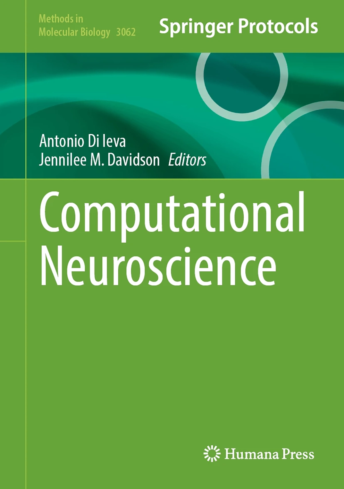
Computational Neuroscience
Di Ieva, A., Davidson, J. (Eds)(2026). Computational neuroscience. Springer. https://link.springer.com/book/9781071654392
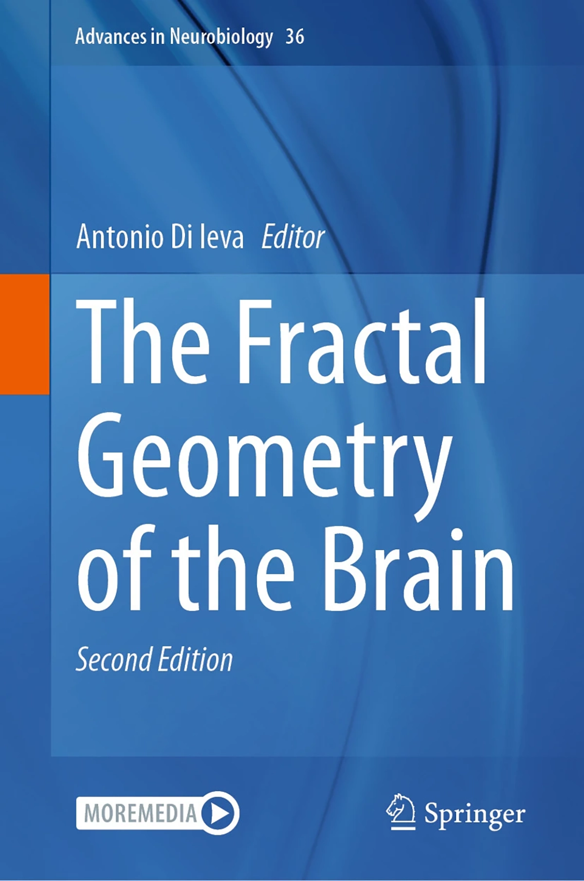
The Fractal Geometry of the Brain
Di Ieva, A., (2024). The Fractal Geometry of the Brain. Springer. https://link.springer.com/book/10.1007/978-3-031-47606-8
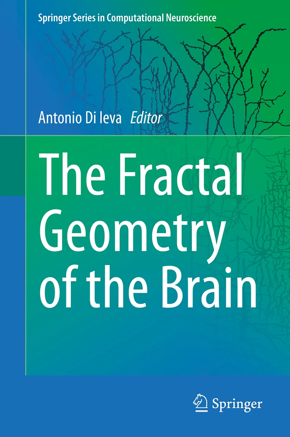
The Fractal Geometry of the Brain
Di Ieva A. (2016). The Fractal Geometry of the Brain. Springer. https://link.springer.com/book/10.1007/978-1-4939-3995-4?page=2
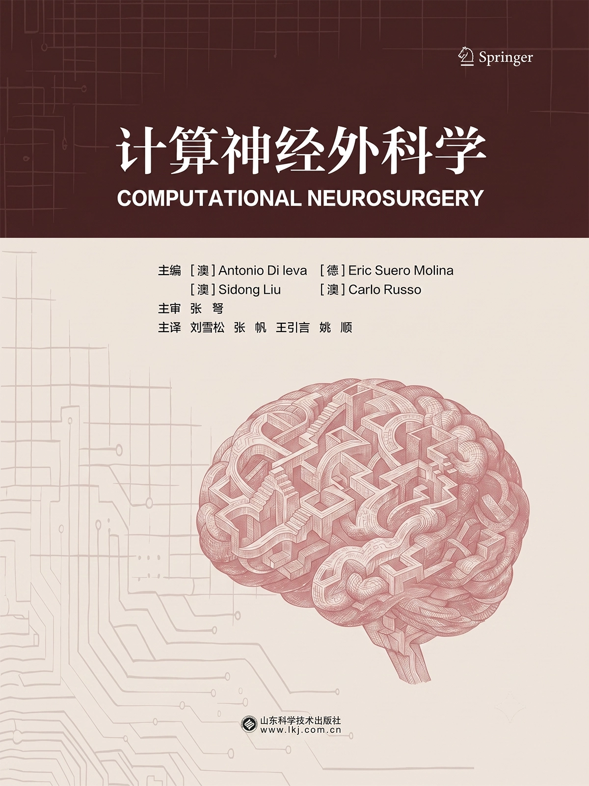
Computational Neurosurgery - Chinese Translation
Di Ieva, A., Suero Molina, E., Liu, S., & Russo, C. (Eds.) (2026). Computational neurosurgery. Springer.
Computational Pipelines for Protein Ubiquitylation Analysis and Prediction
Potapenko, A. et al. (2026). Computational Pipelines for Protein Ubiquitylation Analysis and Prediction. In: Zhang, X., Chen, R., Li, J. (eds) Protein Post-Translational Modifications. Methods in Molecular Biology, vol 3018. Humana, New York, NY. https://doi.org/10.1007/978-1-0716-5166-7_4
The fractal geometry of the brain: an overview
Di Ieva A.. The fractal geometry of the brain: an overview. The fractal geometry of the brain. 2024;:3--13.
Neurosurgery, explainable AI, and legal liability
Matulionyte R., Suero Molina E., Di Ieva A.. Neurosurgery, explainable AI, and legal liability. Computational neurosurgery. 2024;:543--553.
Meta-transfer learning for brain tumor segmentation: within and beyond glioma
Yan S., Liu S., Di Ieva A., Pagnucco M., Song Y.. Meta-transfer learning for brain tumor segmentation: within and beyond glioma. Computational neurosurgery. 2024;:221--230.
Machine and deep learning in hyperspectral fluorescence-guided brain tumor surgery
Suero Molina E., Black D., Xie A., Gill J., Di Ieva A., Stummer W.. Machine and deep learning in hyperspectral fluorescence-guided brain tumor surgery. Computational neurosurgery. 2024;:245--264.
Large language models in neurosurgery
Di Ieva A., Stewart C., Suero Molina E.. Large language models in neurosurgery. Computational neurosurgery. 2024;:177--198.
Fractals, pattern recognition, memetics, and AI: a personal journal in the computational neurosurgery
Di Ieva A.. Fractals, pattern recognition, memetics, and AI: a personal journal in the computational neurosurgery. The fractal geometry of the brain. 2024;:273--283.
Fractals in the neurosciences: a translational geographical approach
Andronache I., Peptenatu D., Ahammer H., Radulovic M., Djuričić G.J., Jelinek H.F., Russo C., Di Ieva A.. Fractals in the neurosciences: a translational geographical approach. The fractal geometry of the brain. 2024;:953--981.
Fractals in neuroimaging
Lahmiri S., Boukadoum M., Di Ieva A.. Fractals in neuroimaging. The fractal geometry of the brain. 2024;:429--444.
Fractals in neuroanatomy and basic neurosciences: an overview
Di Ieva A.. Fractals in neuroanatomy and basic neurosciences: an overview. The fractal geometry of the brain. 2024;:141--147.
Fractal-based analysis of arteriovenous malformations (AVMs)
Di Ieva A., Reishofer G.. Fractal-based analysis of arteriovenous malformations (AVMs). The fractal geometry of the brain. 2024;:413--428.
Fractal-based analysis of histological features of brain tumors
Al-Kadi O.S., Di Ieva A.. Fractal-based analysis of histological features of brain tumors. The fractal geometry of the brain. 2024;:501--524.
Fractal time series: background, estimation methods, and performances
Porcaro C., Moaveninejad S., D’Onofrio V., DiIeva A.. Fractal time series: background, estimation methods, and performances. The fractal geometry of the brain. 2024;:95--137.
Fractal geometry meets computational intelligence: future perspectives
Livi L., Sadeghian A., Di Ieva A.. Fractal geometry meets computational intelligence: future perspectives. The fractal geometry of the brain. 2024;:983--997.
Fractal dimension studies of the brain shape in aging and neurodegenerative diseases
Davidson J.M., Zhang L., Yue G.H., Di Ieva A.. Fractal dimension studies of the brain shape in aging and neurodegenerative diseases. The fractal geometry of the brain. 2024;:329--363.
Fractal dimension analysis in neurological disorders: an overview
Díaz Beltrán L., Madan C.R., Finke C., Krohn S., Di Ieva A., Esteban F.J.. Fractal dimension analysis in neurological disorders: an overview. The fractal geometry of the brain. 2024;:313--328.
Fractal analysis in clinical neurosciences: an overview
Di Ieva A.. Fractal analysis in clinical neurosciences: an overview. The fractal geometry of the brain. 2024;:261--271.
Declaration of computational neurosurgery
Di Ieva A., Suero Molina E., Somerville M.A., Beheshti A., Staartjes V.E., Serra C., Theodore N., Elliott J.M., Wesselink E.O., Russo C., Pilitsis J.G., Bennett C.C., Wu S., Hammond F.M., Lozano A.M., Cusimano M.D., Davidson J.M., Castellano J.F., Okonkwo D.O., Arefan D., Lee C., Zanier O., Da Mutten R., Matula C., Rutka J.T., Pease M., Liu S., Stummer W., Matulionyte R., Yang H., Yuwen C., Cheng X., Fan H., Wang X., Ge Z., Cepeda S., Sheehan J.P., Yang J.Y.M., Hamer R.P., Cohen-Gadol A., Hansford J.R., Savage G., Sowman P.F., Stewart C., Kateb B., Sherif C., Perperidis A., Guller A., Hanft S., D’Amico R.S., Sav A., Cong C., Song Y., Nicolosi F., Wiedmann M.K.H., Barone D.G., Noorani I., Magnussen J., Krieg S.M., Meling T.R., De Ridder D., Lawton M.T., Rosenfeld J.V.. Declaration of computational neurosurgery. Computational neurosurgery. 2024;:11--20.
Cross-Modality Synthesis of T1c MRI from Non-contrast Images Using GANs: Implications for Brain Tumor Research
Tabassum M., Rana P., Suero Molina E., Di Ieva A., Liu S.. Cross-Modality Synthesis of T1c MRI from Non-contrast Images Using GANs: Implications for Brain Tumor Research. Artificial Intelligence in Medicine. 2024;:60–69.
Computational neurosurgery: Foundation
Di Ieva A., Suero Molina E., Liu S., Russo C.. Computational neurosurgery: Foundation. Computational neurosurgery. 2024;:1--8.
Computational fractal-based neurosurgery
Di Ieva A., Davidson J.M., Russo C.. Computational fractal-based neurosurgery. Computational neurosurgery. 2024;:97--105.
Computational fractal-based analysis of MR susceptibility-weighted imaging (SWI) in neuro-oncology and neurotraumatology
Di Ieva A.. Computational fractal-based analysis of MR susceptibility-weighted imaging (SWI) in neuro-oncology and neurotraumatology. The fractal geometry of the brain. 2024;:445--468.
Computational and translational fractal-based analysis in the translational neurosciences: an overview
Di Ieva A.. Computational and translational fractal-based analysis in the translational neurosciences: an overview. The fractal geometry of the brain. 2024;:781--793.
Computational fractal-based analysis of brain tumor microvascular networks
Di Ieva A., Al-Kadi O.S.. Computational fractal-based analysis of brain tumor microvascular networks. The fractal geometry of the brain. 2024;:525--544.
Artificial intelligence, radiomics, and computational modeling in skull base surgery
Suero Molina E., Di Ieva A.. Artificial intelligence, radiomics, and computational modeling in skull base surgery. Computational neurosurgery. 2024;:265--283.
Artificial intelligence in brain tumors
Suero Molina E., Azemi G., Russo C., Liu S., Di Ieva A.. Artificial intelligence in brain tumors. Computational neurosurgery. 2024;:201--220.
Artificial intelligence methods
Liu S., Russo C., Suero Molina E., Di Ieva A.. Artificial intelligence methods. Computational neurosurgery. 2024;:21--38.
Analyzing eye paths using fractals
Newport R.A., Liu S., Di Ieva A.. Analyzing eye paths using fractals. The fractal geometry of the brain. 2024;:827--848.
An experimental verification of neurophysics treatment by executing a multifractal analysis of surface electromyography signals on trapezius, abdominals, and adductor muscles in athletes
Rinzivillo C., Kaleagasioglu F., Casciaro F., Scoppa F., Marvulli R., Ware K., Conte E., Di Ieva A.. An experimental verification of neurophysics treatment by executing a multifractal analysis of surface electromyography signals on trapezius, abdominals, and adductor muscles in athletes. Complexity science in human change. 2024;:81--117.
Robotics for approaches to the anterior cranial fossa
Anokwute M.C., Christodoulides A., Campbell R.G., Harvey R.J., Ieva A.D.. Robotics for approaches to the anterior cranial fossa. Robotics in skull-base surgery. 2023;:35--52.
Publications
Generative adversarial networks in digital pathology and histopathological image processing: a review
Digital pathology is gaining prominence among the researchers with developments in advanced imaging modalities and new technologies. Generative adversarial networks (GANs) are a recent development in the field of artificial intelligence and since their inception, have boosted considerable interest in digital pathology. GANs and their extensions have opened several ways to tackle many challenging histopathological image processing problems such as color normalization, virtual staining, ink removal, image enhancement, automatic feature extraction, segmentation of nuclei, domain adaptation and data augmentation. This paper reviews recent advances in histopathological image processing using GANs with special emphasis on the future perspectives related to the use of such a technique. The papers included in this review were retrieved by conducting a keyword search on Google Scholar and manually selecting the papers on the subject of H&E stained digital pathology images for histopathological image processing. In the first part, we describe recent literature that use GANs in various image preprocessing tasks such as stain normalization, virtual staining, image enhancement, ink removal, and data augmentation. In the second part, we describe literature that use GANs for image analysis, such as nuclei detection, segmentation, and feature extraction. This review illustrates the role of GANs in digital pathology with the objective to trigger new research on the application of generative models in future research in digital pathology informatics.
Computational Neurosurgery in brain tumors: a paradigm shift on the use of artificial intelligence and connectomics in pre- and intra-operative imaging
Computational Neurosurgery is a novel field where computational modeling and artificial intelligence (AI) are used to analyze diseases of neurosurgical interest. Our aim is to apply AI models to brain tumor (BT) images to a) automatically segment BTs on pre-operative MRI, b) predict the genetic subtype of glioma on intra- and post-operative histological specimens; and c) predict the extent of resection according to connectomics data. For the segmentation task, we used 510 BT images to train a deep learning (DL) model for automatic segmentation of the tumors’ edges and comparison of the AI-generated masks versus experts’ consensus (quantified by means of the dice score). For the histopathology task, we digitalized 266 hematoxylin/eosin slides of gliomas (including 130 IDH-wildtype and 136 IDH-mutant) and applied a DL architecture to predict the IDH genetic status, then validated by immunohistochemistry and genetic sequencing. The datasets were also augmented by generating synthetic glioma images by means of a Generative Adversarial Network methodology. The resection of 10 BTs was also customized according to connectomics data. In the segmentation experiment, we reached a dice score of ~0.9 (out of 1.0), while further demonstrating that only the T1, T1 after gadolinium, and FLAIR sequences are necessary for accurate automatic segmentation. In the histopathology task, we were able to predict the genetic status with accuracy between 76% and 95% using the DL model. The machine learning-based connectome analysis allowed us to perform safe supramaximal resection. We have shown the robustness of applying AI methodology or the automatic segmentation of BTs in MR imaging. Moreover, we have also shown that AI can be used to predict the genetic status, specifically, IDH, in histopathology images of gliomas. Our results support the use of AI in the clinical scenario for a fast and objective computerized characterization of patients affected by BTs.
Brain Volumetric and Fractal Analysis of Synthetic MRI: a comparative study with conventional 3D T1-weighted images
Purpose The estimation of brain volumetric measurements based on Synthetic MRI (SyMRI) is easy and fast, however, the consistency of brain volumetric and morphologic measurements based on SyMRI and 3D T1WI should be further addressed. The current study evaluated the impact of spatial resolution on brain volumetric and morphologic measurements using SyMRI, and test whether the brain measurements derived from SyMRI were consistent with those resulted from 3D T1WI. Method Brain volumetric and fractal analysis were applied to thirty healthy subjects, each underwent four SyMRI acquisitions with different spatial resolutions (1 × 1 × 2 mm, 1 × 1x3mm, 1 × 1 × 4 mm, 2 × 2 × 2 mm) and a 3D T1WI (1 × 1 × 1 mm isotropic). The consistency of the SyMRI measurements was tested using one-way non-parametric Kruskal-Wallis test and post hoc Dwass-Steel-Critchlow-Fligner test. The association between SyMRI and 3D T1WI derived measurements was evaluated using linear regression models. Results Our results demonstrated that both in- and through-plane resolutions show an impact on brain volumetric measurements, while brain parenchymal volume showed high consistency across the SyMRI acquisitions, and high association with the measurements from 3D T1WI. In addition, SyMRI with 1 × 1 × 4 mm resolution showed the strongest association with 3D T1WI compared to other SyMRI acquisitions in both volumetric and fractal analyses. Moreover, substantial differences were found in fractal dimension of both gray and white matter between the SyMRI and 3D T1WI tissue segmentations. Conclusions Our results suggested that the measurements from SyMRI with relatively higher in-plane and lower through-plane resolution (1 × 1 × 4 mm) are much closer to 3D T1WI.
Application of deep learning for automatic segmentation of brain tumors on magnetic resonance imaging: a heuristic approach in the clinical scenario
Purpose
Accurate brain tumor segmentation on magnetic resonance imaging (MRI) has wide-ranging applications such as radiosurgery planning. Advances in artificial intelligence, especially deep learning (DL), allow development of automatic segmentation that overcome the labor-intensive and operator-dependent manual segmentation. We aimed to evaluate the accuracy of the top-performing DL model from the 2018 Brain Tumor Segmentation (BraTS) challenge, the impact of missing MRI sequences, and whether a model trained on gliomas can accurately segment other brain tumor types.
Methods
We trained the model using Medical Decathlon dataset, applied it to the BraTS 2019 glioma dataset, and developed additional models using individual and multimodal MRI sequences. The Dice score was calculated to assess the model’s accuracy compared to ground truth labels by neuroradiologists on BraTS dataset. The model was then applied to a local dataset of 105 brain tumors, performance of which was qualitatively evaluated.
Results
The DL model using pre- and post-gadolinium contrast T1 and T2 FLAIR sequences performed best, with a Dice score 0.878 for whole tumor, 0.732 tumor core, and 0.699 active tumor. Lack of T1 or T2 sequences did not significantly degrade performance, but FLAIR and T1C were important contributors. All segmentations performed by the model in the local dataset, including non-glioma cases, were considered accurate by a pool of specialists.
Conclusion
The DL model could use available MRI sequences to optimize glioma segmentation and adopt transfer learning to segment non-glioma tumors, thereby serving as a useful tool to improve treatment planning and personalized surveillance of patients.
Radiomics in gliomas: clinical implications of computational modeling and fractal-based analysis
Radiomics is an emerging field that involves extraction and quantification of features from medical images. These data can be mined through computational analysis and models to identify predictive image biomarkers that characterize intra-tumoral dynamics throughout the course of treatment. This is particularly difficult in gliomas, where heterogeneity has been well established at a molecular level as well as visually in conventional imaging. Thus, acquiring clinically useful features remains difficult due to temporal variations in tumor dynamics. Identifying surrogate biomarkers through radiomics may provide a non-invasive means of characterizing biologic activities of gliomas. We present an extensive literature review of radiomics-based analysis, with a particular focus on computational modeling, machine learning, and fractal-based analysis in improving differential diagnosis and predicting clinical outcomes. Novel strategies in extracting quantitative features, segmentation methods, and their clinical applications are producing promising results. Moreover, we provide a detailed summary of the morphometric parameters that have so far been proposed as a means of quantifying imaging characteristics of gliomas. Newly emerging radiomic techniques via machine learning and fractal-based analyses holds considerable potential for improving diagnostic and prognostic accuracy of gliomas. Key points • Radiomic features can be mined through computational analysis to produce quantitative imaging biomarkers that characterize intra-tumoral dynamics throughout the course of treatment. • Surrogate image biomarkers identified through radiomics could enable a non-invasive means of characterizing biologic activities of gliomas. • With novel analytic algorithms, quantification of morphological or sub-regional tumor features to predict survival outcomes is producing promising results. • Quantifying intra-tumoral heterogeneity may improve grading and molecular sub-classifications of gliomas. • Computational fractal-based analysis of gliomas allows geometrical evaluation of tumor irregularities and complexity, leading to novel techniques for tumor segmentation, grading, and therapeutic monitoring.
Magnetic Resonance Spectroscopic Assessment of Isocitrate Dehydrogenase Status in~Gliomas: The New Frontiers of Spectrobiopsy in Neurodiagnostics
Background
In the era of integrated genomic-histologic analysis of brain tumors, new biomarkers have been introduced as diagnostic, prognostic, and therapeutic indicators. The analysis of the mutation in the isocitrate dehydrogenase (IDH) genes IDH1 and IDH2 has provided important diagnostic and prognostic information for patients affected by diffuse glioma (i.e., the presence of the mutation has been related to an increased survival rate). The reference standard of IDH mutation detection has been its assessment in surgical specimens, immunohistochemistry, and/or genetic sequencing. Knowing the IDH status information preoperatively would be of great importance, because it has been related to tumor progression and the response to treatment. The oncometabolite 2-hydroxyglutarate (2HG), accumulated in gliomas with IDH mutation status, can be detected in vivo using magnetic resonance spectroscopy (MRS).
Methods
The 2HG-MRS technique remains technically challenging. We have summarized the results of the first pilot study in Australia, which included 10 patients affected by glioma. The data recorded from May 2017 to November 2018 were analyzed.
Results
In our exploratory study, we reached a sensitivity and specificity of 100%, confirming the strong predictive role of 2HG, as detected using MRS, in the diagnosis of glioma.
Conclusion
In the present study, we have focused on methodological tips and future perspectives of the technique in the neuroimaging and neuro-oncological scenario. We would advocate the integration of 2HG-MRS into standard clinical practice.
Isocitrate dehydrogenase (IDH) status prediction in histopathology images of gliomas using deep learning
Mutations in isocitrate dehydrogenase genes IDH1 and IDH2 are frequently found in diffuse and anaplastic astrocytic and oligodendroglial tumours as well as in secondary glioblastomas. As IDH is a very important prognostic, diagnostic and therapeutic biomarker for glioma, it is of paramount importance to determine its mutational status. The haematoxylin and eosin (H&E) staining is a valuable tool in precision oncology as it guides histopathology-based diagnosis and proceeding patient’s treatment. However, H&E staining alone does not determine the IDH mutational status of a tumour. Deep learning methods applied to MRI data have been demonstrated to be a useful tool in IDH status prediction, however the effectiveness of deep learning on H&E slides in the clinical setting has not been investigated so far. Furthermore, the performance of deep learning methods in medical imaging has been practically limited by small sample sizes currently available. Here we propose a data augmentation method based on the Generative Adversarial Networks (GAN) deep learning methodology, to improve the prediction performance of IDH mutational status using H&E slides. The H&E slides were acquired from 266 grade II-IV glioma patients from a mixture of public and private databases, including 130 IDH-wildtype and 136 IDH-mutant patients. A baseline deep learning model without data augmentation achieved an accuracy of 0.794 (AUC = 0.920). With GAN-based data augmentation, the accuracy of the IDH mutational status prediction was improved to 0.853 (AUC = 0.927) when the 3,000 GAN generated training samples were added to the original training set (24,000 samples). By integrating also patients’ age into the model, the accuracy improved further to 0.882 (AUC = 0.931). Our findings show that deep learning methodology, enhanced by GAN data augmentation, can support physicians in gliomas’ IDH status prediction.
Deep learning for automated cerebral aneurysm detection on computed tomography images
Purpose: Cerebrovascular aneurysms are being observed with rapidly increasing incidence. Therefore, tools are needed for accurate and efficient detection of aneurysms. We used deep learning techniques with CT angiography acquired from multiple medical centers and different machines to develop and evaluate an automatic detection model. Methods: In this study, we have introduced a deep learning model, the faster RCNN model, in order to develop a tool for automatic detection of aneurysms from medical images. The inputs of the model were 2D nearby projection (NP) images from 3D CTA, which were made by the NP method proposed in this study. This method made aneurysms clearly visible on images and improved the model’s performance. The study included 311 patients with 352 aneurysms, selected from three hospitals, and 208 and 103 of these patients, respectively, were randomly selected to train and test the models. Results: The sensitivity of the trained model was 91.8%. For aneurysm sizes larger than 3 mm, the sensitivity of successful aneurysm detection was 96.7%. We achieved state-of-the-art sensitivity for > 3 mm aneurysms. The sensitivities also indicated that there was no significant difference among aneurysms at different locations in the body. Computing time for the detection process was less than 25 s per case. Conclusions: We successfully developed a deep learning model that can automatically detect aneurysms. The model performed well for aneurysms of different sizes or in different locations. This finding indicates that the deep learning model has the potential to vastly improve clinician performance by providing automated aneurysm detection.
Advanced computational and statistical multiparametric analysis of Susceptibility-Weighted Imaging to characterize gliomas and brain metastases
Susceptibility-weighted imaging (SWI) is a technique useful for evaluation of the internal structures of brain tumors, including microvasculature and microbleeds. Intratumoral patterns of magnetic susceptibility can be quantified by means of fractal analysis. Here, we propose a radiomics methodological pipeline to merge advanced fractal-based computational modelling with statistical analysis to objectively characterize the fingerprint of gliomas and brain metastases. Forty-seven patients with glioma (grades II-IV, according to the WHO 2016 classification system) and fourteen with brain metastases underwent 3 Tesla MRI using a SWI protocol. All images underwent computational analysis aimed to quantify three Euclidean parameters (related to tumor and SWI volume) and five fractal-based parameters (related to the pixel distribution and geometrical complexity of the SWI patterns). Principal components analysis, linear and quadratic discriminant analysis, K-nearest neighbor and support vector machine methods were used to discriminate between tumor types. The combination of parameters offered an objective evaluation of the SWI pattern in gliomas and brain metastases. The model accurately predicted 88% of glioblastoma, according to the quantification of intratumoral SWI features, failing to discriminate the other types. SWI is not normally used to classify brain tumors, however fractal-based multi-parametric computational analysis can be used to characterize intratumoral SWI patterns to objectively quantify tumors-related features. Specific parameters still have to be identified to provide completely automatic computerized differential diagnosis.
A Deep Learning Methodology for Differentiating Glioma Recurrence from Radiation Necrosis using Multimodal MRI: Algorithm Development and Validation (Preprint)
Background: The radiological differential diagnosis between tumor recurrence and radiation-induced necrosis (ie, pseudoprogression) is of paramount importance in the management of glioma patients.
Objective: This research aims to develop a deep learning methodology for automated differentiation of tumor recurrence from radiation necrosis based on routine magnetic resonance imaging (MRI) scans.
Methods: In this retrospective study, 146 patients who underwent radiation therapy after glioma resection and presented with suspected recurrent lesions at the follow-up MRI examination were selected for analysis. Routine MRI scans were acquired from each patient, including T1, T2, and gadolinium-contrast-enhanced T1 sequences. Of those cases, 96 (65.8%) were confirmed as glioma recurrence on postsurgical pathological examination, while 50 (34.2%) were diagnosed as necrosis. A light-weighted deep neural network (DNN)(ie, efficient radionecrosis neural network [ERN-Net]) was proposed to learn radiological features of gliomas and necrosis from MRI scans. Sensitivity, specificity, accuracy, and area under the curve (AUC) were used to evaluate performance of the model in both image-wise and subject-wise classifications. Preoperative diagnostic performance of the model was also compared to that of the state-of-the-art DNN models and five experienced neurosurgeons.
Results: DNN models based on multimodal MRI outperformed single-modal models. ERN-Net achieved the highest AUC in both image-wise (0.915) and subject-wise (0.958) classification tasks. The evaluated DNN models achieved an average sensitivity of 0.947 (SD 0.033), specificity of 0.817 (SD 0.075), and accuracy of 0.903 (SD 0.026), which were significantly better than the tested neurosurgeons (P=. 02 in sensitivity and P<. 001 in specificity and accuracy).
Conclusions: Deep learning offers a useful computational tool for the differential diagnosis between recurrent gliomas and necrosis. The proposed ERN-Net model, a simple and effective DNN model, achieved excellent performance on routine MRI scans and showed a high clinical applicability.
medinform.jmir.org
Letter to the Editor Regarding The Exoscope in Neurosurgery: An Innovative Point of View. A Systematic Review of the Technical, Surgical, and Educational Aspects
How I do it: 3D exoscopic endoscope-assisted microvascular decompression
Background
Microvascular decompression (MVD) is an effective treatment for drug-resistant trigeminal neuralgia and hemifacial spasm. However, failure of symptomatic improvement can arise from difficulties in identifying and/or decompressing the offending vessel. Microscopic and endoscopic techniques have been used to improve visualisation and safety of the procedure but there are limitations to each technique.
Method
A 3D exoscopic endoscope-assisted MVD technique is described, including advice on potential pitfalls.
Conclusion
Compared with the standard microscope-assisted techniques, the 3D exoscopic endoscope-assisted MVD offers an improved visualisation without compromising the field of view within and outside the surgical field.
Computational quantitative MR image features - a potential useful tool in differentiating glioblastoma from solitary brain metastasis
Purpose
Glioblastomas (GBM) and metastases are the most frequent malignant brain tumors in the adult population. Their presentation on conventional MRI is quite similar, but treatment strategy and prognosis are substantially different. Even with advanced MR techniques, in some cases diagnostic uncertainty remains. The main objective of this study was to determine whether fractal, texture, or both MR image analyses could aid in differentiating glioblastoma from solitary brain metastasis.
Method
In a retrospective study of 55 patients (30 glioblastomas and 25 solitary metastases) who underwent T2W/SWI/CET1 MRI, quantitative parameters of fractal and texture analysis were estimated, using box-counting and gray level co-occurrence matrix (GLCM) methods.
Results
All five GLCM parameters obtained from T2W images showed significant difference between glioblastomas and solitary metastases, as well as on CET1 images except correlation (SCOR), contrary to SWI images which showed different values of two parameters (angular second moment-SASM and contrast-SCON). Only three fractal features (binary box dimension-Dbin, normalized box dimension-Dnorm and lacunarity-λ) measured on T2W and Dnorm measured on CET1 images significantly differed GBMs from solitary metastases. The highest sensitivity and specificity were obtained from inverse difference moment (SIDM) on T2W and SIDM on CET1 images, respectively. Combination of several GLCM parameters yielded better results. The processing of T2W images provided the most significantly different parameters between the groups, followed by CET1 and SWI images.
Conclusions
Computational-aided quantitative image analysis may potentially improve diagnostic accuracy. According to our results texture features are more significant than fractal-based features in differentiation glioblastoma from solitary metastasis.
AI-augmented multidisciplinary teams: hype or hope?
Over the past few years, the approach to treating diseases has switched from the solo expertise of a single specialist to treatment orchestrated by a multidisciplinary team, with the intention of merging the opinions of various experts in an optimum way to treat patients. Multidisciplinary teams are the backbone of the decision making that forms the basis of modern medicine. The multi disciplinary team offers a more hierarchical, highlevel, and complex system compared with a single specialist, and aims to determine the most plausible differential diagnosis, relate this to the most probable prognosis, and select the best course of treatment. An incorrect differential diagnosis might lead to error cascades, the consequences of which are inaccurate expectations and imprecise treatments. However, multidisciplinary teams can still be limited by insufficient expertise of the single members, outofdate knowledge on the relevant evidence based medical literature, consolidated teams that are not open to new opinions or participants, and logistical or communication barriers. In recent years, the use of artificial intelligence (AI) in medicine has greatly increased, although the field is still challenged by particular concerns and the absence of validation against the opinions of experts or across different populations1 and by the absence of a standardised method of analysis. 2 AI methods can be used to help clinicians and surgeons with decision making, to reduce errors in judgment and improve differential diagnosis, and thereby improve the choice of treatment and patient outcome. The underlying fear of a dystopic challenge wherein AI is in competition with human experts can be overcome by viewing AI as a means to create a socalled augmented physician, with the exper tise of specialists enhanced by AI. This alternative view would shift the paradigm from one of humanversus machine, to humanandmachine. 3 Machines will not replace physicians, but physicians using AI will soon replace those not using it. Likewise, decisions from the multidisciplinary team meetings of the next generation are likely to be implemented by the machine, as AI will support more objective and reliable decision making, reducing the limitations of human error and subjectivity related to the single specialist or team. As stated by Enrico Coiera, 4 the way to prepare for these coming times is adapting clinical education to the digital world, to build the capacity for enhanced decision making and prognostication among physicians by means of AI. Continuous vigilance around advice validation will still be of paramount importance, 5 to avoid physicians blindly following the machinetracked pathway over the course of a patient’s diagnosis and therapy. In addressing the need to adapt in response to the fate of medicine in this time of AI, 4 the fate of multidisciplinary teams is to change as well.
Transcranial colour duplex and central aortic pressure measurements in the management of cerebral arteriovenous malformations: a pilot study using non-invasive measures
Background: Following removal of an arteriovenous malformation of the brain (bAVM) the redistribution of blood can impose several clinically challenging issues including intracranial haemorrhage and arteriovenous capillary hypertensive syndrome. The underlying mechanism of such complications remains controversial, although control of blood pressure has been recognised as an integral component in haemorrhage prevention. Serial daily non-invasive monitoring for patients in this instance would be beneficial in improving management. Transcranial colour duplex (TCD) is a potential technique for providing pressure measurements and real-time, dynamic, haemodynamic spectra. Aim: To establish whether blood outflow velocity in the middle cerebral vein (MCV) can be quantified in the days following bAVM resection and whether values differ from other types of intracranial surgery. Methods: Blood pressure and TCD of 13 patients (aged 46±19 y, 7 female) having bAVMs resected and 7 patients having other intracranial surgeries (control group, aged 48±15 y, 6 female) were studied for days 1 to 3 following surgery. Ultrasound via the transtemporal window was used to obtain diameter, as well as peak and end-diastolic velocity of the MCV. Brachial blood pressure was also obtained using an automatic oscillometric blood pressure monitor. Results: Systolic (bAVM 96±2 mmHg, control 89±10 mmHg; P=0.68) and diastolic blood pressure (bAVM 55±2 mmHg, control 48±7 mmHg; P=0.92) did not differ between the groups. MCV peak systolic velocity was greater in the bAVM group (34±20 cm/s, controls 20±9 cm/s; P=0.049). The control group had peak systolic velocities ranging from 9 to 32 cm/s. Peak systolic velocity in the bAVM group varied from 9 to 98 cm/s. End diastolic velocity (bAVM 13±11 cm/s, control 13±6 cm/s; P=0.89) and diameters (bAVM 37±19 mm, control 26±7 mm; P=0.16) did not differ between the groups. Two of the bAVM patients that had sustained high MCV peak velocities had a post-operative haemorrhage. Conclusions: Unusually elevated blood flow velocities and diameters were observed in the MCV of patients following bAVM resection. Findings on this small data set provide insight into plausible vessel remodelling and elucidate a post-operative time frame when vessels may have impaired autoregulation of cerebral blood flow.
The Fractal Geometry of the Brain
Reviews the most intriguing applications of fractal analysis in neuroscience with a focus on current and future potential, limits, advantages, and disadvantages. Will bring an understanding of fractals to clinicians and researchers also if they do not have a mathematical background, and will serve as a good tool for teaching the translational applications of computational models to students and scholars of different disciplines. This comprehensive collection is organized in four parts: (1) Basics of fractal analysis; (2) Applications of fractals to the basic neurosciences; (3) Applications of fractals to the clinical neurosciences; (4) Analysis software, modeling and methodology.