Neuroimaging
Computational Neuroimaging
Reading the brain and its tumours from images alone.
Overview
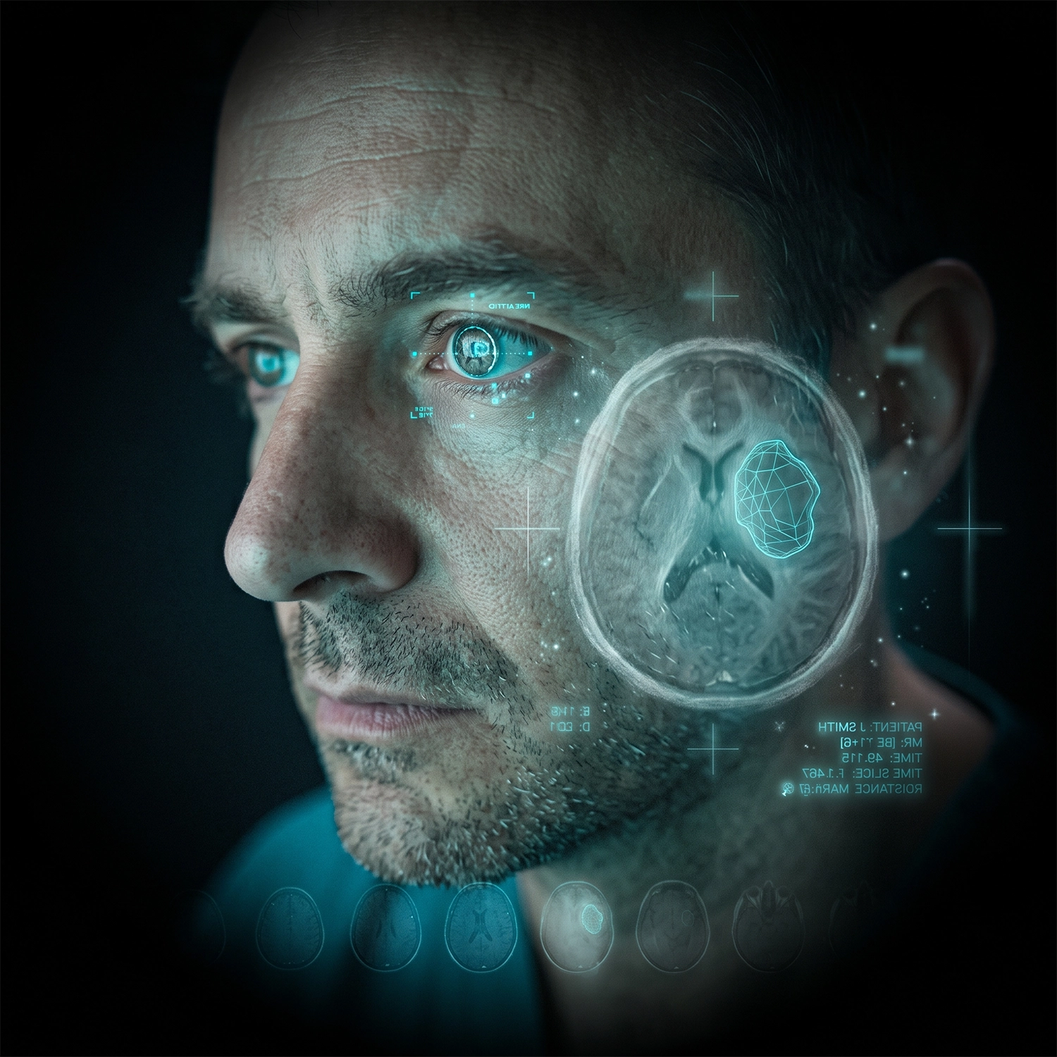
Computational Neuroimaging is where the lab turns clinical scans into quantitative, decision-ready information. We develop and validate methods that analyse MRI, PET and spectroscopic data to segment, classify and grade brain tumours, and increasingly to read molecular and genetic signatures directly from imaging, non-invasively, before a scalpel is ever involved. A central thread of our work is radiomics and radiogenomics: extracting high-dimensional quantitative features from routine scans and linking them to underlying tumour biology, so that imaging can predict molecular subtype, aggressiveness and likely treatment response. Building on this, the lab pioneered the spectrobiopsy concept in Australia, the idea that spectroscopic imaging can act as a virtual biopsy, characterising tissue at the molecular level without surgical sampling. Underpinning all of this is a commitment to methods that are robust, reproducible and clinically translatable. We build standardised acquisition and analysis pipelines, deep-learning models for segmentation and grading, and validation across multi-institutional cohorts, so that what works in a paper has a path to working at the bedside.
Methods
Techniques & approaches
The computational methods that underpin this research area
Radiomics
Publications
Selected publications in this area

Journal article
2024
Use of artificial intelligence in the prediction of Chiari malformation type 1 recurrence after posterior fossa decompressive surgery
King V., Liu S., Russo C., Jayasekara M., Stoodley M., Di Ieva A.
Cureus

Review
2024
Radiomics and machine learning in brain tumors and their habitat: a systematic review
Tabassum M., Suman A.A., Suero-Molina E., Pan E., Di Ieva A., Liu S.
Cancers
Journal article
2024
Computational Neurosurgery in brain tumors: a paradigm shift on the use of artificial intelligence and connectomics in pre- and intra-operative imaging
Di Ieva A., Russo C., Suman A.A., Liu S.
Neuro-Oncology
Book chapter
2024
Fractals in neuroimaging
Lahmiri S., Boukadoum M., Di Ieva A.
The fractal geometry of the brain
Funding
Selected funding in this area
Grants supporting our computational neuroimaging research programme
NHMRC
AI in brain tumour imaging: towards augmented diagnostics
$1,556,663 (+$311,332 MQ)
Ideas Grant
Australian Research Council
In search of relevant things: a novel approach for image analysis
$1,015,000
Future Fellowship
Tour de Cure
RAGE radiogenomics in paediatric tumours
$100,000
Research Grant
Macquarie University
Computational modelling & AI in brain tumour neuroimaging
$20,000
Safety Net Scheme
Browse publications.
Join our work.
Explore the full body of CNS Lab research or contact us to discuss collaboration opportunities.