Microscopic
Computational Digital Neuropathology
Quantifying disease from the tissue up.
Overview
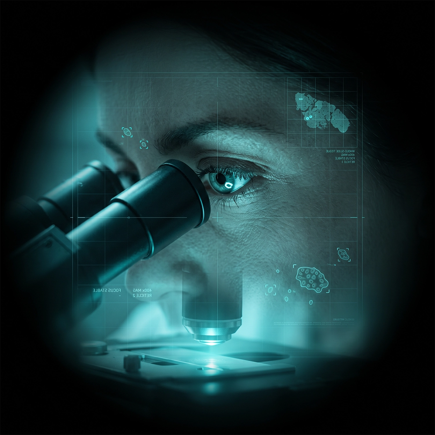
Computational Digital Neuropathology brings the lab's quantitative approach to the microscope. Working with whole-slide images of brain tissue, we develop deep-learning and pathomics methods that detect, classify and characterise disease at the cellular and tissue level, turning what has traditionally been a qualitative, expert-driven read into reproducible, quantitative information. Our work includes automated analysis of tumour histology, extraction of pathomic features that capture cell morphology and tissue architecture, and models that link these features to diagnosis, grade and molecular status. A recurring theme is collaboration across institutions and modalities, bridging digital pathology with imaging and genomics so that a tumour can be understood across scales, from the whole brain down to the single cell. Because pathology sits at the heart of diagnosis, we place particular emphasis on robustness and generalisability: models that hold up across scanners, stains and sites, and that clinicians can trust.
Methods
Techniques & approaches
The computational methods that underpin this research area
Radiomics
Publications
Selected publications in this area

Conference paper
2024
TPMIL: Trainable Prototype enhanced Multiple Instance Learning for whole slide image classification
Yang L., Mehta D., Liu S., Mahapatra D., Di Ieva A., Ge Z.
Medical Imaging with Deep Learning 2023

Journal article
2024
Combination Drug Therapy of Glioblastoma: Lessons from 3D In Vitro Models and the Roadmap for Future Research
Vaezzadeh M., Kachooei E., Krishnamurthy S., Manandhar P., Nadort A., Guillemin G.J., Di Ieva A., Santiago M., Heng B., Guller A.
Advanced Therapeutics
Book chapter
2024
Fractal dimension analysis in neurological disorders: an overview
Díaz Beltrán L., Madan C.R., Finke C., Krohn S., Di Ieva A., Esteban F.J.
The fractal geometry of the brain
Journal article
2024
Deep learning-based hyperspectral image correction and unmixing for brain tumor surgery
Black D., Gill J., Xie A., Liquet B., Di leva A., Stummer W., Suero Molina E.
iScience
Funding
Selected funding in this area
Grants supporting our computational neuroimaging research programme
Australian Cancer Research Foundation (ACRF)
COMET Centre for Advanced Cancer Modelling & Experimental Oncology
$2,500,000
Centre Grant
Browse publications.
Join our work.
Explore the full body of CNS Lab research or contact us to discuss collaboration opportunities.